Download
1 / 48
480 likes | 645 Vues
Acute Pancreatitis, its management and Complications A Case Study. By Ayesha Aslam and Ambreen Qayyum. scenario.

E N D
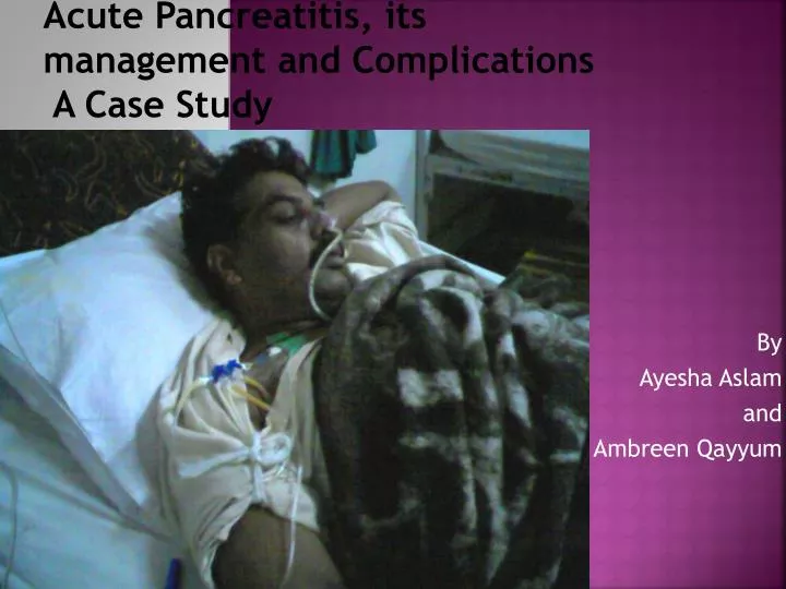
Acute Pancreatitis, its management and Complications A Case Study By Ayesha Aslam and Ambreen Qayyum
scenario A 45 years old male presented with pain in upper abdomen and on and off vomiting for 2 months. Pain was sudden, severe, continuous and not relieved by medication. He also complained of dark colored urine.
OnGPE • Patient appears as if in pain • Slightly dyspneic • NG intubation • CV line maintained • Jaundiced • No edema, lymphadenopathy
On abdominal examination • On inspection: Slow abdominal movements • On palpation: Tense tender abdomen Tenderness and guarding in upper abdomen Vague mass in epigastrium • On percussion: Percussion painful Dull in epigastrium • On auscultation: Bowel sounds not audible
Investigations • Laboratory investigations: serum amylase ……………….. serum bilirubin ……………... BSR…………………………………… serum alkaline phosphate.. • USG abdomen: Pancreas showing heterogeneous texture with a few necrotic areas • CT scan: Pancreatic head and body tissue is almost completely replaced by hemorrhagic or exudative fluid
Acute Necrotizing Pancreatitis with Pseudo cyst Formation diagnosis
Physiology • Secretions • Enzymes • Hormones • Bicarbonates
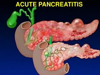
Acute Pancreatitis Overview • Inflammation of the pancreas • Non infectious • Autodigestive process • Proenzymes converted to enzymes • lysosomal and zymogen granule compartments fuse, enabling activation of trypsinogen to trypsin • intracellular trypsin triggers the entire zymogen activation cascade • secretory vesicles extruded across the membrane into the interstitium • molecular fragments act as chemo attractants for inflammatory cells. • Activated neutrophils exacerbate the problem by releasing super oxides or proteolytic enzymes • macrophages release cytokines that further mediate local (and, in severe cases, systemic) inflammatory responses • early mediators- tumor necrosis factor-alpha, interleukin-6, and interleukin-8.
Causes of Acute Pancreatitis • Obstruction • Choledocholithiasis (gallstone) • Toxins and medications • Ethanol • Scorpion venom • Organophosphate pesticides • Medications • Trauma • Abdominal trauma • Endoscopic retrograde cholangiopancreatography
Causes Of Acute Pancreatitis Metabolic derangements Hypercalcemia Hypertriglyceridemia Infections Parasitic Viral--mumps, hepatitis A, varicella, cytomegalovirus, human immunodeficiency virus Bacterial--mycoplasma, legionella, M. tuberculosis Vascular Ischemia Embolic Vasculitis Idiopathic
Initial Presentation • PAIN (cardinal symptom) • Epigasrtium • May be sudden or gradual • Mild to severe • Often radiating to back • Typically dull, constant • worse when lying flat • Triggered by ingestion of large meal or alcohol
CONT… • Other symptoms • Nausea • Vomiting • Fever, chills or both • Features of dehydration • Low B.P • Rapid heartbeat (internal bleeding or as a response to pain) • Weakness or feeling tired • Fatigue • Irritability • Confusion • Circulatory Shock
Mild vs. severe attack • Ranson's Criteria for Pancreatitis mortality • Present on Admission: • Age greater than 55 years • WBC greater than 16,000/ul • Blood glucose greater than 200 mg/dl • Serum LDH greater than 350 I.U./L • SGOT (AST) greater than 250 I.U./L
Cont…. • Developing During the First 48 Hours: • Hematocrit fall greater than 10% • BUN increase greater than 8 mg/dl • Serum calcium less than 8 mg/dl • Arterial oxygen saturation less than 60 mm Hg • Base deficit greater than 4 m eq/L • Estimated fluid sequestration greater than 6000 ml (6 liters)
Cont…. • Ranson score of 0-2, minimal mortality • Ranson score of 3-5, 10%-20% mortality • Ranson score of >5 has more than 50% mortality and is associated with more systemic complications.
Glasgow Score for Pancreatitis • age >55 years • pO2 <8.0 kPa • WCC >15x109/litre • Ca2= (uncorr.) <2.0 mmol/L • ALT >100 IU • LDH>600 IU • glucose>10 mmol/L • urea>16 mmol/L • albumin<32g/L
Glasgow Score for Pancreatitis • based on nine biochemical and physiological parameters • Now modified be removal of levels of transaminases • The new score called modified Glasgow score is based on eight parameters • designed for predicting the prognosis of patient with acute Pancreatitis • Each parameters scores 1 point. • A score of 3 or more predicts, but does not define, a severe episode of acute Pancreatitis
Diagnosis • History • Examination • Lab Investigations • Pancreatic, liver and kidney functions • X-ray films • USG Abdomen • CT Scan Abdomen • ERCP
Treatment • Self care at home • Stop alcohol • Diet improvement • OTC medication • Medical care • Objective- relieve symptoms and stop progression • Admit to hospital • Maintenance of oxygenation • Maintenance of IV line • Medication for pain and nausea • Antibiotics in certain settings • Bowel rest by NPO • Nasogastric intubation • Nutritional supplementation
Cont…. • Surgery • Never preferred in uncomplicated cases • Only done to remove the etiological factor e- g cholecystectomy in case of gall stones • Early ERCP and sphincterectomy with stone extraction • In complicated cases surgical procedures required as per consultation
Potential Complications of Acute Pancreatitis • Local complications • Pancreatic necrosis • Pancreatic duct disruption • Pancreatic and peripancreatic fluid accumulation • Pancreatic pseudo cyst formation • Psudoaneurysm formation
Potential Complications of Acute Pancreatitis • Systemic complications • Hypovolemia • Pancreatic necrosis • Extra pancreatic necrosis • Acute respiratory distress syndrome • Acute renal failure • Adynamic ileus • Circulatory shock • Sepsis • Information from Beger HG, Rau B, Mayer J, Pralle U. Natural course of acute Pancreatitis. World J Surg 1997;21:130- 5.
Pancreatic necrosis • In 20% of all cases of Pancreatitis • Cell death with resultant devitalized tissue likely to become infected • Potential outcomes include • Resolution • Psudocyst formation • Abscess
Pseudo cyst formation • Peripancreatic fluid in fibrinous capsule • Rupture can cause peritonitis, fistula formation, erosion of a vessel leading to hemorrhage • Pancreatic ascites or pleural effusion • Hollow viscus obstruction by compression of colon, duodenum, stomach, cbd
Pancreatic abscess • Forms through various mechanisms • secondary infection of a psuedocyst • Collection of pus resulting from necrosis, liquefaction or infarction • Late complication of acute necrotizing Pancreatitis • Most common micro organisms are enterics and Candida sp.
Symptoms indicating a complication • Diagnosis with long course • Hemodynamic instability • Failure of medical therapy • Fluid collection on CT scan • Pain with mass in epigastric region • Abnormal vitals consistent with sepsis • Cullen’s sign • Grey turner’s sign
Management of complications • Medical care • Supportive only • Special attention to B.P and volume status • Antibiotic therapy against enterics
Non-Surgical Management of complications 1.Radiological Percutaneous drainage (USG or CT guided) 2.Endoscopic : stenting for disrupture of pancreatic duct Drainage- Tran gastric, transampullary Advantages 1.Avoid risk of surgery 2.Safe in expert hands 3.Local anesthesia Complications 1.Infection 2.Haemorrhage 3.Displacement of catheter 4.Trauma to viscera 5.fistula formation 6.recurrence
Surgical Management of complications • External drainage • Pancreatic necrosectomy • Indications • 1.Multiple collections • 2.Large collections • 3.Inaccessible sites for percutaneous methods • 4.Infected collections with thick fluid • 5.Failure of percutaneous drainage • 7.Associated with necrosis
Complications of surgery • 1.Prolonged length of hospital stay • 2.Morbidity associated with surgery • 3.Reoperations
Prognosis of fluid accumulations • Most resolve spontaneously • Percutaneous approach better in poor surg risk patients with small collections • In necrosis- percutaneous drainage followed by laparoscopic evacuation of debris • Surgery - backup management • Early diagnosis, clinical vigilance in detecting complications, referral of patients to specialized centers before they become morbid, ensures state of art multidisciplinary management and hastens outcome in acute Pancreatitis
Criteria for Multiple Organ System Failure (MOSF) • Cardiovascular • Mean arterial pressure <=50 mm Hg. • Need for volume loading and/or vasoactive drugs to maintain systolic blood pressure above 100 mm Hg. • Heart rate <=50 beats per minute. • Ventricular tachycardia/fibrillation. • Cardiac arrest. • Acute myocardial infarction. • Pulmonary • Respiratory rate <=5 per minute or >=50 per minute. • Mechanical ventilation for 3 or more days • Renal • Serum creatinine >=3.5 mg per dL (280 mmol per L). • Dialysis/ ultra filtration needed.
Cont…. • Neurological • Glasgow Coma Scale ¾ 6 (in the absence of sedation). • Hematological • Hematocrit <=20%. • Leukocyte count <=300 per mm3 (0.3 3 109 per L). • Platelet count <=50 3 103 per mL (50 3 109 per L). • Disseminated intravascular coagulation. • Hepatic • Total bilirubin level >=3.5 mg per dL (51 mmol per L) in the absence of hemolysis. • ALT >100 U per L. • Gastrointestinal • Stress ulcer necessitating transfusion of more than 2 units of blood per 24 hours. • Acalculus cholecystitis. • Necrotizing enterocolitis. • The sum of the failing organ system during a day. Score varies from zero to 7.