Download
1 / 34
350 likes | 596 Vues
BRAINSTEM I Neuroscience 2005. LECTURE OVERVIEW. Definition of Brainstem Brief Gross Orientation Brief Cross Sectional Anatomy Orientation Correlation of Gross and Cross Sectional Anatomy Composite List of Brainstem Structures Cranial Nerves (CN’s) Introduction Nucleus vs. Ganglion

E N D
LECTURE OVERVIEW • Definition of Brainstem • Brief Gross Orientation • Brief Cross Sectional Anatomy Orientation • Correlation of Gross and Cross Sectional Anatomy • Composite List of Brainstem Structures • Cranial Nerves (CN’s) • Introduction • Nucleus vs. Ganglion • Organization / Classification Based on Embryology • Spinal Cord vs. Brainstem • Skeletal mm: Branchial Arch vs. Somite Myotome • Correlation of Location and Function of CN’s
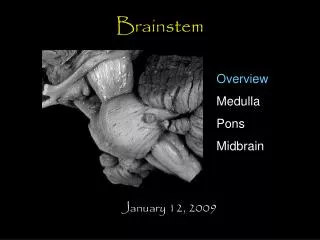
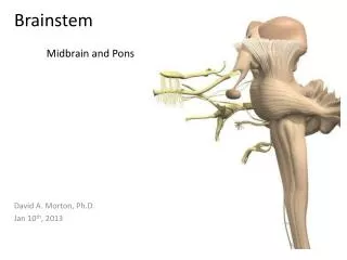
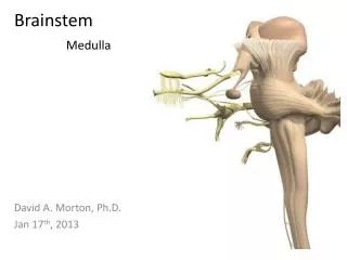
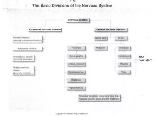
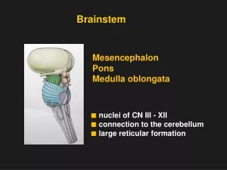
DIVISIONS of the BRAINSTEM • Midbrain • Pons • Medulla (Terms “upper” and “lower” sometimes used)
LOCATION of BRAINSTEM • Cranial Cavity • Begins caudally at FORAMEN MAGNUM • Mostly infratentorial • CNS • Between spinal cord and diencephalon Carpenter, Core Text of Neuroanatomy, 1991
Brainstem in situ Nolte Text, p. 263
Median Saggital Plane Haines Atlas Haines Atlas Nolte Text, p. 267
SERIAL SECTIONS Haines Atlas
CLINCAL IMAGING vs. ANATOMY WHICH WAY IS UP? VENTRAL DORSAL Haines Atlas
CLASSICAL REPRESENTATIVE SECTIONS MEDULLA PONS MIDBRAIN Haines Atlas
Nolte Fig. 15-3 • Brainstem Structures: • Name • Location • Function • Dev. Divisions • Mesen • Meten • Myelen
Cranial Nerves Telencephalon 1 (not pictured) Diencephalon 2 Midbrain 3 & 4 Pons 5 - 8 Medulla 9 - 12 Spinal Cord 11
Cranial Nerves (CN’s) • HEAD AND NECK INNERVATION • Components of CN’s: • Sensory • Special sensory • Motor (2 embryological types of skeletal mm) • Autonomic Motor: Parasympathetics (sm. mm, glds) - preganglionic - head to the transverse colon - where do the sympathetics to the head originate? • Neurons of origin in: - NUCLEI - GANGLIA • Clinical relevance: - localization of lesions - extent of damage / prognosis - difficult to treat
WITHIN CNS CLUSTER OF NEURONS NOT PSEUDOUNIPOLAR* SENSORY OR MOTOR OUTSIDE OF CNS CLUSTER OF NEURONS PSEUDOUNIPOLAR SENSORY CRANIAL NERVENUCLEUS vs. GANGLION *except mesencephalic nucleus of V Carpenter, Core Text of Neuroanatomy, 1991
SENSORY GANGLIA of CRANIAL NERVES Langman’s Embryology Carpenter, Core Text of Neuroanatomy, 1991
“WORM DIAGRAMS” NOLTE HAINES ATLAS
CLASSIFICATION OF CRANIAL NERVES • Based on: A. Function • Motor • Somatic (skel. mm) • Visceral (sm. & card. mm & spec. skel. mm) • Sensory • Special Sensory B. Embryologically Derived Location • Alar plate = sensory (lateral) • Basal plate = motor (medial)
SENSORY and MOTORORGANIZATION Embryonic CNS Spinal Cord Brainstem Langman’s Embryology
Motoneurons (EFF) : somatic motor (skel. mm) visceral motor (sm. & card. mm., glands) Sensory neurons (AFF): somatic sensory (skin, muscle, muc. mem., bone, and joint receptors) visceral sensory (viscera) Motoneurons (EFF): somatic motor (skel. mm) visceral motor (sm. &card. mm., glands) branchial motor(skel. mm) Sensory neurons (AFF): somatic sensory ( skin, muscle., muc. mem., bone, and joint receptors) visceral sensory (viscera) special visceral(taste, smell) special somatic(eye, ear) *vagus and accessory nn. also to body SPINAL CORD (body) BRAINSTEM (head*)
Somatic vs. Branchiomeric Skeletal Muscle • Somatic: from the myotomes (part of somites) • extraocular mm = CN III, IV, VI • tongue mm = CN XII • Branchial: from branchial arch* musculature • mm of mastication = CN V • mm of facial expression = CN VII • mm of the pharynx and larynx = CN’s IX, X, XI (ambiguus) *also referred to as “pharyngeal arch”
Pharyngeal Arches • Pharyngeal = Visceralso skel. musc. from arches is a “special” kind of “visceral” muscle (SVE) • The term Pharyngeal Arch is used interchangeably with Branchial Arch • BRANCHIAL motor = SVE Langman’s Embryology
BRANCHIAL ARCHES (PHARYNGEAL ARCHES) Langman’s Embryology 1st = V musc. mast., ant. belly dig., mylohy., tensor tym., tensor palat. 2nd = VII musc. facial expr., stapedius, post. belly dig., stylohyoid, auricular 3rd = IX stylopharyngeus 4th = X Sup. Laryngeal Br. = cricothyroid, levator palat., pharyng. constr. ( 6th arch supplied by Recurrent Laryngeal Br. = intrinsic laryn. mm; also CN XI)
Langman’s Embryology Nolte Text, p. 376 Gilman & Winans-Newman
Special Sensory Somatic Sensory Visceral Sensory Visceral motor Branchial motor Somatic motor Nolte Fig. 12-1
G 5, 7, 9, 10 = skin + mucous mem. (prop. 3, 4, 6, 12) S S 2, 8 = eye + ear A G 9, 10 = int. organs V S 7, 9, 10, 1 = taste, olf. S G 12, 3, 4, 6 = tongue + eye mm (myotomes) E G 3, 7, 9, 10 = parasymp. V S 5, 7 ambig. = branchial mm