Download
1 / 67
670 likes | 675 Vues
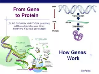
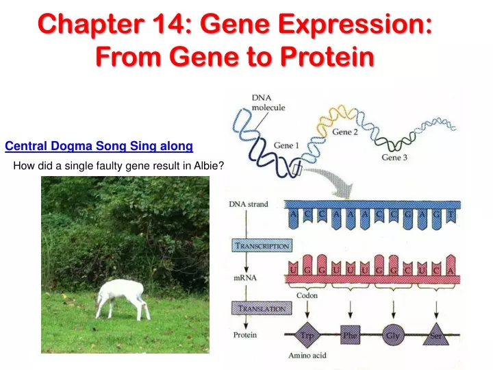
Chapter 14: Gene Expression: From Gene to Protein. Central Dogma Song Sing along. How did a single faulty gene result in Albie?. What is the function of DNA?. The information content of DNA is in the form of specific sequences of nucleotides along the DNA strands

E N D
Chapter 14: Gene Expression: From Gene to Protein Central Dogma Song Sing along How did a single faulty gene result in Albie?
What is the function of DNA? • The information content of DNA • is in the form of specific sequences of nucleotides along the DNA strands • It codes for the production of proteins • Aka: Protein Synthesis
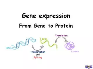
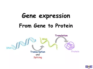
The DNA inherited by an organism • leads to specific traits by dictating the synthesis of proteins • The process by which DNA directs Protein Synthesis, Gene expression • includes two stages: • Transcription • Translation
Where does this process take place? • The ribosome • Is part of the cellular machinery for translation, polypeptide synthesis in all living organisms Figure 17.1: A model of a ribosome
Evidence from the Study of Metabolic Defects • In 1909, British physician Sir Archibald Garrod • Was the first to suggest that genes dictate phenotypes through enzymes that catalyze specific chemical reactions in the cell • An inheritable disease result from the inability to produce a certain enzyme • He referred to certain diseases as caused by “inborn errors of metabolism”
Nutritional Mutants in Neurospora: Scientific Inquiry • Beadle and Tatum causes bread mold to mutate with X-rays • Creating mutants that could not survive on minimal medium • Disabled genes one by one & looked for changes in each mutant’s phenotype thus seeing the normal function of the gene • Preview
EXPERIMENT RESULTS Class I Mutants Class II Mutants Class III Mutants Wild type Minimal medium (MM) (control) MM + Ornithine MM + Citrulline MM + Arginine (control) Working with the mold Neurospora crassa, George Beadle and Edward Tatum had isolated mutants requiring arginine in their growth medium and had shown genetically that these mutants fell into three classes, each defective in a different gene. From other considerations, they suspected that the metabolic pathway of arginine biosynthesis included the precursors ornithine and citrulline. Their most famous experiment, shown here, tested both their one gene–one enzyme hypothesis and their postulated arginine pathway. In this experiment, they grew their three classes of mutants under the four different conditions shown in the Results section below. • Using genetic crosses • they determined that their mutants fell into three classes, each mutated in a different gene The wild-type strain required only the minimal medium for growth. The three classes of mutants had different growth requirements Figure 17.2
From the growth patterns of the mutants, Beadle and Tatum deduced that each mutant was unable to carry out one step in the pathway for synthesizing arginine, presumably because it lacked the necessary enzyme. Because each of their mutants was mutated in a single gene, they concluded that each mutated gene must normally dictate the production of one enzyme. Their results supported the one gene–one enzyme hypothesis and also confirmed the arginine pathway. (Notice that a mutant can grow only if supplied with a compound made after the defective step.) CONCLUSION Class I Mutants (mutation in gene A) Class II Mutants (mutation in gene B) Class III Mutants (mutation in gene C) Wild type Precursor Precursor Precursor Precursor Enzyme A Gene A A A A Ornithine Ornithine Ornithine Ornithine Enzyme B Gene B B B B Citrulline Citrulline Citrulline Citrulline Enzyme C Gene C C C C Arginine Arginine Arginine Arginine George Beadle speaks to you
Beadle and Tatum developed the: • “one gene–one enzyme hypothesis” • Which states that the function of a gene is to dictate the production of a specific enzyme • As researchers learned more about proteins • they made minor revision to the one gene–one enzyme hypothesis • Genes code for polypeptide chains or for RNA molecules which code for the production of enzymes
Basic Principles of Transcription and Translation • Transcription • Is the synthesis of messenger RNA (mRNA) under the direction of DNA • Translation • Is the actual synthesis of a polypeptide, which occurs under the direction of mRNA • Occurs onribosomesof all cells
DNA TRANSCRIPTION RNA polymerase mRNA DNA Ribosome mRNA TRANSLATION Polyribosome Direction of transcription 0.25 m RNA polymerase Polypeptide DNA (a) Prokaryotic cell. In a cell lacking a nucleus, mRNAproduced by transcription is immediately translatedwithout additional processing. Polyribosome Polypeptide (amino end) Ribosome mRNA (5 end) • In prokaryotes • Transcription and translation occur together (no nuclear envelope) Prokaryotic Transcription & Translation occurs simultaneously Figure 17.3a
Nuclear envelope DNA TRANSCRIPTION Pre-mRNA RNA PROCESSING mRNA Ribosome TRANSLATION (b) Eukaryotic cell. The nucleus provides a separatecompartment for transcription. The original RNAtranscript, called pre-mRNA, is processed in various ways before leaving the nucleus as mRNA. Polypeptide Figure 17.3b • In eukaryotes • RNA transcripts are modified before becoming true mRNA DNA – Pre-mRNA – mRNA - Polypeptide
Cells are governed by a cellular chain of command DNARNA Protein Francis Crick's “Central Dogma” of molecular biology: “DNA makes RNA makes protein.” This general rule emphasized the order of events from transcription through translation history.nih.gov/exhibits/nirenberg/glossary.htm
The Genetic Code The order of the nucleotides (Nitrogen bases) codes for the genetic code How many bases correspond to an amino acid? If only 2 bases in a codon, there would only be 16 codons (42) • Is encoded as a sequence of nonoverlapping base triplets, or codons • 43 = 64
Gene 2 DNA molecule Gene 1 Gene 3 DNA strand (template) 5 3 A C C T A A A C C G A G TRANSCRIPTION A U C G C U G G G U U U 5 mRNA 3 Codon TRANSLATION Gly Phe Ser Trp Protein Figure 17.4 Amino acid • During transcription • The gene determines the sequence of bases along the length of an mRNA molecule
Second mRNA base U C A G U UAU UUU UCU UGU Tyr Cys Phe UAC UUC UCC UGC C U Ser UUA UCA UAA Stop Stop UGA A Leu UAG UUG UCG Stop UGG Trp G CUU CCU U CAU CGU His CUC CCC CAC CGC C C Arg Pro Leu CUA CCA CAA CGA A Gln CUG CCG CAG CGG G Third mRNA base (3 end) First mRNA base (5 end) U AUU ACU AAU AGU Asn Ser C lle AUC ACC AAC AGC A Thr A AUA ACA AAA AGA Lys Arg Met or start G AUG ACG AAG AGG U GUU GCU GAU GGU Asp C GUC GCC GAC GGC G Val Ala Gly GUA GCA GAA GGA A Glu Figure 17.5 GUG GCG GAG GGG G Cracking the Code • A codon in messenger RNA • Is either translated into an amino acid or serves as a translational stop signal
The Genetic Code In the Genetic Code, there are 43 = 64 (# of N-bases# of bases in a codon) possible codons
Evolution of the Genetic Code • The genetic code is nearly universal • Shared by organisms from the simplest bacteria to the most complex animals • UUU in bacteria & a blue whale codes for Phenylalanine
In laboratory experiments • Genes can be transcribed and translated after being transplanted from one species to another • All three organisms below are expressing a firefly gene inserted into their genome Figure 17.6
Molecular Components of Transcription • RNA synthesis • Is catalyzed by RNA polymerase, which pries the DNA strands apart and hooks together the RNA nucleotides Follows the same base-pairing rules as DNA, except that in RNA, Uracil substitutes for Thymine
3 1 2 Promoter Transcription unit 5 3 3 5 Start point DNA RNA polymerase Initiation. After RNA polymerase binds to the promoter, the DNA strands unwind, and the polymerase initiates RNA synthesis at the start point on the template strand. Template strand of DNA 5 3 3 5 Unwound DNA RNA transcript Elongation. The polymerase moves downstream, unwinding the DNA (3` to 5`) and elongating the RNA transcript 5 3 . In the wake of transcription, the DNA strands re-form a double helix. Rewound RNA 5 3 3 5 3 RNA transcript 5 Termination. Eventually, the RNA transcript is released, and the polymerase detaches from the DNA. 5 3 3 5 3 5 Completed RNA transcript Figure 17.7 Synthesis of an RNA Transcript • The stages of transcription are • Initiation • Elongation • Termination
General Initiation of Transcription 3` to 5` on DNA 5` to 3` for mRNA
Eukaryotic promoters 1 TRANSCRIPTION DNA Pre-mRNA RNA PROCESSING mRNA Ribosome TRANSLATION Polypeptide Promoter 5 3 A T A T A A A A T A T T T T 3 5 TATA box Start point Template DNA strand Several transcription factors 2 Transcription factors 5 3 3 5 Additional transcription factors 3 RNA polymerase II Transcription factors 3 5 5 3 5 RNA transcript Figure 17.8 Transcription initiation complex RNA Polymerase Binding andInitiation of Transcription • Promoters signal the initiation of RNA synthesis • Series of T’s & A’s (TATA box) going in a 3` to 5` direction on DNA • Transcription factors • Help eukaryotic RNA polymerase recognize promoter sequences • Complex of transcription factors and RNA polymerase II bound to the promoter is called a transcription initiation complex. UTR UTR UTR
Elongation Non-template strand of DNA RNA nucleotides RNA polymerase T A C C A T A T C 3 U 3 end T G A U G G A G E A C C C A 5 A A T A G G T T Direction of transcription (“downstream”) 5 Template strand of DNA Newly made RNA
Elongation of the RNA Strand • As RNA polymerase moves along the DNA • it continues to untwist the double helix, exposing about 10 to 20 DNA bases at a time for pairing with RNA nucleotides • A single gene can be transcribed simultaneously by several molecules of RNA polymerase following each other.
The mechanisms of termination • are different in prokaryotes and eukaryotes • Eukaryotic cells modify RNA after transcription • Enzymes in the eukaryotic nucleus modify pre-mRNA or the primary transcript in specific ways before the genetic messages are dispatched to the cytoplasm
A modified guanine nucleotide added to the 5 end 50 to 250 adenine nucleotides added to the 3 end TRANSCRIPTION DNA Polyadenylation signal Protein-coding segment Pre-mRNA RNA PROCESSING 5 3 mRNA G P P AAA…AAA P AAUAAA Ribosome Start codon Stop codon TRANSLATION Poly-A tail 5 Cap 5 UTR 3 UTR (Untranslated region) Polypeptide Alteration of mRNA Ends • Each end of a pre-mRNAmolecule is modified in a particular way • The 5end receives a modified nucleotide cap (GTP cap – Guanine triphosphate) • The 3end gets a Poly-A tail • Facilitates the export of mature mRNA from the nucleus • Protects mRNA from breakdown by hydrolytic enzymes • Helps ribosomesattache to the 5` end of mRNA in the cytoplasm • UTR are untranslated regions Figure 17.9
Exon Intron 5 Exon Intron Exon 3 5 Cap Poly-A tail Pre-mRNA TRANSCRIPTION DNA 30 31 104 105 146 1 Pre-mRNA RNA PROCESSING Introns cut out and exons spliced together Coding segment mRNA Ribosome TRANSLATION 5 Cap Poly-A tail mRNA Polypeptide 1 146 3 UTR 3 UTR Split Genes and RNA Splicing • Average gene ~ 1,200 nucleotides (400 Amino Acids) • ~ length of transcription unit is 8,000 nucleotides therefore it needs to be cut down. • Most eukaryotic genes have long noncoding stretches of nucleotides, introns, between coding sections, exons • RNA splicing • Removes intronsand joins exons (coding regions) • Animation Figure 17.10
Role of Splicesomes - animation • Is carried out by spliceosomes in some cases • Made up of Small nuclear ribonucleic acid (snRNA) w/ specific proteins Figure 17.11
Ribozymes • Ribozymes • Are catalytic RNA molecules that function as enzymes and can splice rRNA • Pre-rRNA actually can remove its own introns.
Gene DNA Exon 1 Exon 2 Intron Exon 3 Intron Transcription RNA processing Translation Domain 3 Domain 2 Domain 1 Polypeptide • Proteins often have a modular architecture • consisting of discrete structural and functional regions called domains • In many cases • different exons code for the different domains in a protein • By the removal of introns between the domains, the polypeptide can then be produced
Many genes can give rise to two or more polypeptides depending on which segments are treated as exons during RNA processing – Alternate RNA splicing.
Why Introns and Split Genes? • Though proteins synthesized in a linear fashion, they are actually modular w/ various functional regions called domains • Allows for additional crossing over of chromosomes • Genes of our evolutionary past from prokaryotic organisms (they don’t have introns) • Promoters & terminal signals for turning on and off genes
Molecular Components of Translation • A cell translates a mRNA message into protein- polypeptide • With the help of transfer RNA (tRNA)
DNA TRANSCRIPTION mRNA Ribosome TRANSLATION Polypeptide Amino acids Polypeptide tRNA with amino acid attached Ribosome Trp Phe Gly tRNA C C C G G Anticodon A A A A G G G U G U U U C Codons 5 3 mRNA Translation: the basic concept Figure 17.13
tRNA & anticodon Amino Acid tRNA Anticodon Actually a single RNA strand ~ 80 nucleotides long with nitrogenous bases H bonding to each other causing the strand to fold on itself into a cloverleaf
3 A Amino acid attachment site C C 5 A C G C G C G U G U A A U U A U C G * G U A C A C A * A U C C * G * U G U Figure 17.14a G G * G A C C G * C A G * U G * * G A G C Hydrogen bonds (a) G Two-dimensional structure. The four base-paired regions and three loops are characteristic of all tRNAs, as is the base sequence of the amino acid attachment site at the 3 end. The anticodon triplet is unique to each tRNA type. (The asterisks mark bases that have been chemically modified, a characteristic of tRNA.) C U A G * A * A C * U A G A Anticodon The Structure and Function of Transfer RNA A • Molecules of tRNA are not all identical (45 total) • Each carries a specific amino acid on one end • Each has an anticodon on the other end • A tRNA molecule • Consists of a single RNA strand that is only about 80 nucleotides long • Is roughly L-shaped (or t) C
Wobble w/ 3rd anticodon.U can bond with A or G and still not change the coded amino acid (AGA & AGG codes for arginine
Amino acid attachment site 5 3 Hydrogen bonds A A G 3 5 Anticodon Anticodon (c) Symbol used in this book (b) Three-dimensional structure tRNA’s three dimensional structure Figure 17.14b
A specific enzyme called anaminoacyl-tRNAsynthetase • joins each amino acid to the correct tRNA • There is one for each amino acid Figure 17.15
Ribosomes • Facilitate the specific coupling of tRNA anticodons with mRNA codons during protein synthesis • mRNA bonds first to the small subunit of the ribosomes then the large subunit attaches
DNA TRANSCRIPTION mRNA Ribosome TRANSLATION Polypeptide Exit tunnel Growing polypeptide tRNA molecules Large subunit E P A Small subunit 5 3 mRNA (a) Computer model of functioning ribosome. This is a model of a bacterial ribosome, showing its overall shape. The eukaryotic ribosome is roughly similar. A ribosomal subunit is an aggregate of ribosomal RNA molecules and proteins. • The ribosomal subunits • Are constructed of proteins and RNA molecules named ribosomal RNA or rRNA Figure 17.16a
P site (Peptidyl-tRNA binding site) A site (Aminoacyl- tRNA binding site) E site (Exit site) Large subunit mRNA binding site Small subunit (b) Schematic model showing binding sites. A ribosome has an mRNA binding site and three tRNA binding sites, known as the A, P, and E sites. This schematic ribosome will appear in later diagrams. • The ribosome has three binding sites for tRNA • The P site – primary bonding site • The A site – adjacent bonding site • The E site – exit site E P A Figure 17.16b
Schematic model with mRNA and tRNA. A tRNA fits into a binding site when its anticodon base-pairs with an mRNA codon. The P site holds the tRNA attached to the growing polypeptide. The A site holds the tRNA carrying the next amino acid to be added to the polypeptide chain. Discharged tRNA leaves via the E site through an exit tunnel. **Translation of tRNA is always in the 5` to 3` direction of mRNA **Protein is built in the 3` to 5` direction (like DNA!) (c) 3` 5` Figure 17.16c
Certain antibiotics can inactivate bacterial ribosomes without interferring with eukaryotic ribosomes & normal protein synthesis. • Tetracycline and Streptomycin. • Bacterial ribosomes are smaller than eukaryotic ribosomes.
Building a Polypeptide • We can divide translation into three stages • Initiation • Elongation • Termination