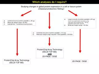
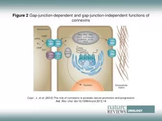
Download
1 / 1
10 likes | 117 Vues
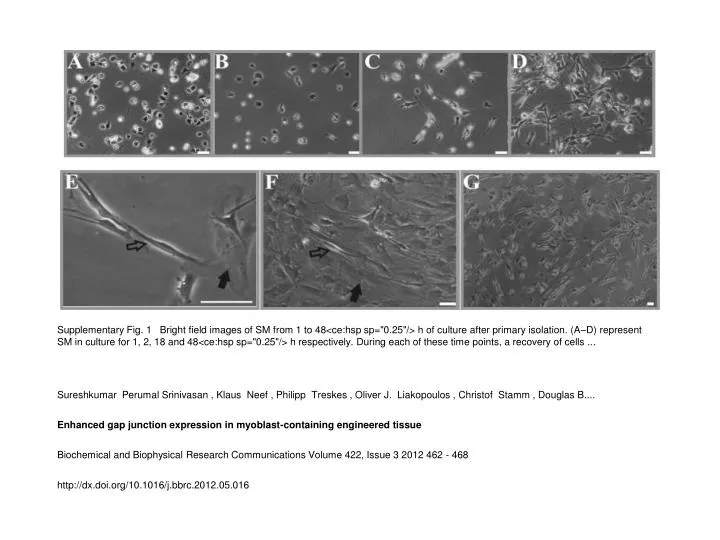
Supplementary Fig. 1 Bright field images of SM from 1 to 48<ce:hsp sp="0.25"/> h of culture after primary isolation. (A–D) represent SM in culture for 1, 2, 18 and 48<ce:hsp sp="0.25"/> h respectively. During each of these time points, a recovery of cells.

E N D
Supplementary Fig. 1 Bright field images of SM from 1 to 48<ce:hsp sp="0.25"/> h of culture after primary isolation. (A–D) represent SM in culture for 1, 2, 18 and 48<ce:hsp sp="0.25"/> h respectively. During each of these time points, a recovery of cells ... Sureshkumar Perumal Srinivasan , Klaus Neef , Philipp Treskes , Oliver J. Liakopoulos , Christof Stamm , Douglas B.... Enhanced gap junction expression in myoblast-containing engineered tissue Biochemical and Biophysical Research Communications Volume 422, Issue 3 2012 462 - 468 http://dx.doi.org/10.1016/j.bbrc.2012.05.016