Download
1 / 12
130 likes | 269 Vues
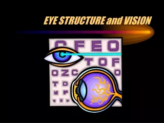
Eye Structure and Seeing Light. The eye is like a camera: Light enters, is focused on a surface, and a picture is made. Light enters your eye through a clear portion of the sclera (the tough, white, outer covering of the eye), called the cornea.

E N D
The eye is like a camera: Light enters, is focused on a surface, and a picture is made. Light enters your eye through a clear portion of the sclera (the tough, white, outer covering of the eye), called the cornea.
The cornea is curved, so it slightly bends the light as it goes through. Light then passes through the aqueous humor (a clear fluid for eye nourishment, in the anterior chamber) and through the pupil. The pupil is simply a hole in the iris.
The iris is a muscle that controls the size of the pupil. The iris is the colored part of the eye. • In bright light, the iris expands and the pupil gets smaller • In low light, the iris contracts and the pupil gets bigger
Directly behind the iris is the lens. This structure changes shape to focus the light so that we can see clearly. Its shape is convex, meaning it curves outward on both sides. The ciliary muscles above and below the lens control the shape of the lens.
Behind the lens is a clear gel called the vitreous humor. After moving through the vitreous humor, the light strikes the retina. The retina is the lining on the inside of the back of the eye that contains two types of light-sensitive cells: rods and cones.
Rods sense black and white and work in low light. Cones sense color and must have more light than rods to work. Three kinds of cones: L-cones sense long wavelengths in the red range M-cones sense mid-range wavelengths in green range S-cones sense short wavelengths in the blue range
The rods and cones send messages to the brain through the optic nerve. The brain makes sense of all the information it is receives. In your brain, the sight center is in the back, between your ears. This location explains why a blow to the back of your head might cause blindness, even though your eyes are fine.
Two Causes of Color Blindness • Genetic(you are born with these types) Sometimes a cone is missing, or the cone does not recognize the correct wavelengths of light. L- and M-cone problems result in red-green color blindness, the most common.
2. Non-Genetic(these types occur after birth) Accidents that damage the vision center of the brain, cataracts, glaucoma, Parkinson’s Disease can cause S-cone problems, diabetic retinopathy can affect color vision
Eye Anatomy Review cornea pupil iris anterior chamber aqueous humor lens vitreous humor retina fovea choroid sclera optic nerve
Image Sources 2004 Microsoft Corporation, One Microsoft Way, Redmond, WA 98052-6399 USA. National Cancer Institute at the National Institutes of Health http://www.cancer.gov/cancertopics/pdq/treatment/retinoblastoma/patient/page1/AllPages/Print Wikipedia http://en.wikipedia.org/wiki/File:Schematic_diagram_of_the_human_eye_en.svg MedLine Plus, National Institutes of Health http://www.nlm.nih.gov/medlineplus/ency/presentations/100206_1.htm MedLine Plus, National Institutes of Health http://www.nlm.nih.gov/medlineplus/ency/imagepages/9482.htm National Eye Institute, National Institutes of Health http://www.nei.nih.gov/healthyeyes/eyeexam.asp Federal Aviation Administration http://www.hf.faa.gov/webtraining/VisualDisplays/HumanVisSys2.htm Federal Aviation Administration http://www.hf.faa.gov/webtraining/visualdisplays/HumanVisSys6.htm Glaucoma, MedLine Plus, National Institutes of Health http://www.nlm.nih.gov/medlineplus/ency/imagepages/9349.htm