Download
1 / 17
170 likes | 264 Vues
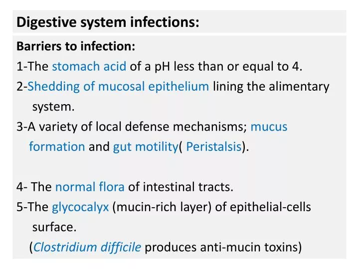
Digestive system infections:. Barriers to infection: 1-The stomach acid of a pH less than or equal to 4. 2- Shedding of mucosal epithelium lining the alimentary system. 3-A variety of local defense mechanisms; mucus formation and gut motility ( Peristalsis ).

E N D
Digestive system infections: Barriers to infection: 1-The stomach acid of a pH less than or equal to 4. 2-Shedding of mucosal epithelium lining the alimentary system. 3-A variety of local defense mechanisms; mucus formation and gut motility( Peristalsis). 4- The normal flora of intestinal tracts. 5-The glycocalyx (mucin-rich layer) of epithelial-cells surface. (Clostridium difficileproduces anti-mucin toxins)
N 6-The Bile salts detergent action. 7-The secreted antimicrobial peptides. (EnterotoxigenicE.coliproduces heat-labile toxin that suppress these peptides. 8-Mcells of Peyer patches have surveillance function. 9-Secretory IgA.
Establishment of infectious diseases in the digestive system: The defense barriers are changed in favor of the microbe due to: 1-Anatomic alterations: A-Obstructionsto the flow of intestinal secretions (gallbladder stones). B-Surgery may create intestinal “blind loops” that are isolated from the moving stream of intestinal contents. Absence of flushing action of intestinal secretions. Bacterial overgrowth syndrome; malabsorption.
N 2-Changes in stomach acidity; due to proton-pump inhibitors: -Decreased pathogenic dose results in colonization of intestinal mucosa; ExampleSalmonella species. -Shigella species and E.coliO157:H7 are acid resistance. 3-Alterations to the normal flora; due to broad-spectrum antibiotics. Pseudomembrane colitis; Clostridium difficile. 4-Invasion of Gut by virulent microbial strains.
Intestinal invasive diseases , inflammation and damage: 1-Invasive Enteritis, and dysentery (bloody diarrhea): A-Shigelladysenteriae infection: Pathogenic dose: less than 200 CFU. Reservoir: human colon only (no animal carriers). Transmission: Fecal-Oral, Person to Person. Pathogenesis: - Endotoxin triggers inflammation. - Shiga toxin type I: Enterotoxic and cytotoxic activities. It is interfering with 60S ribosomal subunit; necrosis. -The microbe invades the M cell in the lumen of Gut.
N Multiply inside these cells, using actin polymerization to infect neighboring epithelial cells. Released , engulfed by intestinal macrophages. Escape from APC, infects other epithelial cells. Very Shallow ulceration of intestinal mucosa.
N Enterocolitis, shigellosis (most severe form is dysentery). Fever , lower abdominal cramps; diarrhea first watery, then bloody with mucus. Invasive infection: shallow ulcerative Enterocolitis. Hemolytic-Uremic syndrome. B-Entamoebahistolytica(dysentery). Microbiology: Gram-negative short bacilli, Nonmotile, Non-spore formers. Enterobacteriaceae grow best on XLD. Facultative anaerobic, non-Lactose fermenters. Can not produce H2S and identify by API 20E
2-Invasive Enteritis and Hemorrhagic Colitis:Enterohemorrhagic E. coli: - Transmission: food (hamburger), milk, Fecal-Oral. - Incubation period: 3-5 days. - Reservoir: Cattle; bovine feces, pork. - Pathogenic dose: 10-100 CFU. - Toxin: Verotoxin : Exotoxin - Pathogenesis: - EHEC bind to cells in the large intestinal mucosa(Caecum,Colon). - Verotoxin-Epithelial receptor interaction. - Decrease protein synthesis by interfering with 60 S ribosomal subunit.
N - Intracellular localization inside lysozyme. - Cell death; apoptosis. - Mucosal necrosis; Hemorrhagic Colitis with bloody diarrhea. - Shiga like exotoxins; Hemolytic-uremic syndrome. Microbiology: -Gram negative short bacilli. -Lactose fermenters. - Metallic green sheen on EMB medium. - Indole positive.
N 3-Invasive Enteritis and Bloody watery diarrhea: A- Campylobacter jejuni infection: Reservoir: intestinal tracts of humans, cattle, sheep, dogs, cats, and Poultry. Transmission: -Fecal-Oral (direct cont.), Ingestion of contaminated meat (poultry), contaminate water, or unpasteurized milk. Incubation period: 3-5 days. Microbiology: Gram-negative curved helical rods with polar flagella.
N - Microaerophilic ( 10% CO2, 5% O2, and 85% N2) microbe with an optima of 42˚C temp. - Humidity should be > 95%. - Resistance for Cephalothin. - Sensitive for Nalidixic acid. - Catalase and oxidase positive.
Pathogenesis of Campylobacter jejuni: Pathogenicdose: as few as 500 CFU. The microbe invades the small and large intestinal mucosa. Colonization of intestinal epithelial layer, engulfed by intestinal dendritic cells(DC). DC release inflammatory mediators, chemotaxis, cellular infiltration. Ulcerative, inflammatory lesions in the jejunum, and ileum. UlcerativeColitis. Pus and RBCs in stool; AcuteEnteritis (common cause of infectious diarrhea worldwide). - Traveler’s diarrhea and pseudo-appendicitis.
N n
B-Salmonella enteriditisand Salmonella typhimurium : Reservoir: -Human: Large intestinal tracts: Carriage state. -Animals: most important: Chicken. Transmission: 1-Fecal-Oral from carrier person. 2-Ingestion of contaminated chicken products (raw chicken, eggs). Incubationperiod: 6-48 hours.
Pathogenesis: TheMicrobe invades the ileocecal region lymphoid tissue. Invasion of Lamina propria; endotoxin activity. Dendritic cell activation; production of TNF and IL-8. PMN cells chemotaxis and PG response. PG stimulate cellular cAMP of epithelial cells. Release of NaCl from intestinal epithelial cells; dehydration. PMN cells prevent mesenteric lymph node and RES invasion. Bloody watery diarrhea. Diagnosis: Clinical: abdominal symptoms and fever. Laboratory: Stool culture: similar to Salmonellatyphi.
C- Listeria monocytogenes: listeriolysin Associated food and Transmission: -Unpasteurized milk products, undercooked meat & raw vegetables -Fresh soft cheese. -Ready-to-eat meat. Incubation period: 8 - 48 hrs. Pathogenesis: Invasion of intestinal epithelium. Intracellular survival: Listeriolysin-O. Cell-mediated immunity. Watery bloody diarrhea. host actin Division Jet formation