Download
1 / 31
370 likes | 1.74k Vues
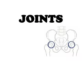
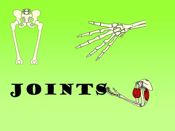
JOINTS. Learning Objectives. To know the different joint types To understand how to classify joint types To be able to discuss the importance of the structure and function of a synovial joint. Joints. The human skeleton is jointed to allow movement.

E N D
Learning Objectives • To know the different joint types • To understand how to classify joint types • To be able to discuss the importance of the structure and function of a synovial joint
Joints • The human skeleton is jointed to allow movement. • Muscular contraction causes the bones to move about the joints. A joint iswhere two or more bones meet andmuscles act together to cause movement.
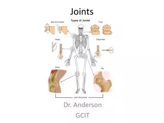
Types of Joints There are 3 main types of joint found in the body. • Fibrous/ immovable joints • The bones at an immoveable joint cannotmove - they overlap or interlock, and are held together by a tough fibre, e.g. theskull. • 2. Cartilaginous/slightly moveable joints • The bones at a slightly moveable joint can only move • a little - they are held together by strong straps • called ligaments and are joined by protective pads • known as cartilage, e.g. theribs. • 3. Synovial joint/freely moveable • At a freely moveable joint the bones move freely. • They are also known as synovialjoints, and • are the largest group of joints found in the body, • e.g. thehips,shouldersandknees.
Freely Moveable Joints • Freely Moveable joints are also known as Synovial Joints. • They are freely moving and occur where 2 or more bones meet. • There are about 70 freely moveable joints in the human skeleton. • These interest us the most because they allow the greatest range of movement and are found in the appendicualr skeleton
Cartilage • Hyaline or Articular cartilage- found on the surface • of bone • 2. Yellow Elastic cartilage- elastic and found in the • external ear • 3. White Fibrocartilage- tough, dense tissue that • acts as a shock absorber. Found between the vertebrae
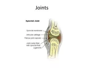
Articular/hyaline Cartilage – A material which covers the end of each bone, and which helps prevent friction between the joint. • Absorbs compression placed on the joint, • protects the joint. • 2. Joint Capsule – The outer layer is a tough fibrous layer called the fibrous capsule. • Increases stability • The inner layer is called the synovial membrane • Secretes synovial fluid, strengthens the joint
3. Synovial Membrane– Thin membrane which lines the inside of the joint capsule. • It produces synovial fluid • 4. Synovial Fluid – The fluid which surrounds the joint and is contained in the joint cavity. • Reduce friction between the cartilage • Nourish the cartilage • Get rid of any waste debris
4. Ligaments – A band of strong fibrous tissue, helps prevent dislocation. • Connect bone to bone • 5. Tendons– Strong connective tissue that attached muscle to bone. • Connect muscle to muscle • 6. Bursa- pad of fat provide cushioning between the fibrous capsule and a bone or muscle. • Cushion the joint and act as shock absorbers
The Synovial Joint of the Knee The knee is a hinge joint. Femur Articular(hyaline)Cartilage Joint capsule Cruciate Ligaments Synovial Membrane Patella Synovial Fluid Joint cavity Tibia/Fibula Tendons
The Synovial Joint of the Hip The Hip is a ball and socket joint. Cartilage Pelvis Synovial Fluid SynovialMembrane Ligaments Tendons Femur
TASK • Label the diagram of a synovial joint using a colour code system • YOU MAY BE ASKED TO LABEL A SYNOVIAL JOINT IN • EXAM OR EXPLAIN THE STRUCTURE AND FUCTION
2. List two features that increase joint stability and give their specific function?
Types of Synovial Joints Synovial joints are classifed according to the shape of the articulating surface. KEY Ball & Socket Joint Hinge Joint Pivot Joint Gliding Joint Saddle Joint Condyloid Joint
KEY TERM ARTICULATION- The different bones that form to make the joint EXAMPLE JointJoint typeArticulating bonesMovement Elbow Hinge joint Humerus, radius, ulna Write down another example
1. Ball and Socket Joints The ball has a head shaped of one bone which articulates with a cuplike socket. Allows the greatest range of movement Examples HIP SHOULDER
2. Hinge Joints They are cylindrical shaped of 1 bone articulates with a depression of an adjacent bone Movement is restricted to bending and straightening EXAMPLES ELBOW KNEE
3. Pivot Joints These are rounded, pointed or concave of one bone which articulates which a ring shaped bone. Movement is restricted to 1 bone rotating around the longitudinal axis. EXAMPLEVERTEBRAE OFTHE NECK
4. Gliding Joints The articulating surfaces are flat. It allows limited movement EXAMPLES HAND BETWEEN THE CARPELS
5. Saddle Joints The articulating surfaces are shaped like a saddle It allows very limited movement EXAMPLE THUMB JOINT
6. Condyloid Joints The surfaces are flatter and oval forming a shallow joint It allows the second greatest range of movement. EXAMPLE WRIST JOINT
Structure and function Hyaline/Articular cartilage Structure: Smooth, spongy cartilage that covers end of bone. Function: prevent friction between bones absorb compression placed on the joint protect bone from getting crushed Joint capsule: Structure: Outer layer is a tough fibrous layer- fibrous membrane.
The inner layer- synovial membrane Function:strengthen the joint secrete synovial fluid Synovial Fluid Structure; slippery fluid, contained in the joint cavity Function; reduce friction between cartilage nourish the articular cartilage get rid of waste debris in joint Ligament Structure; band of strong fibrous tissue Function; connect bone to bone
Meniscus (cartilage) Structure: white fibrocartilage Function: improves the fit between bone ends increases joint stability reduces wear and tear at joint Bursa Structure:fluid filled sacs Function: prevent friction where bones, ligaments and muscles may rub together
PLENARY Identify the bones that articulate at the shoulder joint? (2 marks) Identify the bones that articulate at the elbow joint? (3 marks)
Ball and Socket Joints Examples HIP SHOULDER The ball has a head shaped of one bone which articulates with a cuplike socket. Allows the greatest range of movement Hinge Joints EXAMPLES ELBOW KNEE They are cylindrical shaped of 1 bone articulates with a depression of an adjacent bone Movement is restricted to bending and straightening
Pivot Joints VERTEBRAE OFTHE NECK These are rounded, pointed or concave of one bone which articulates which a ring shaped bone. Movement is restricted to 1 bone rotating around the longitudinal axis. HAND BETWEEN THE CARPELS Gliding Joints The articulating surfaces are flat. It allows limited movement
Saddle Joints EXAMPLE THUMB JOINT The articulating surfaces are shaped like a saddle It allows very limited movement Condyloid Joints The surfaces are flatter and oval forming a shallow joint It allows the second greatest range of movement. EXAMPLE WRIST JOINT