Download
1 / 35
350 likes | 917 Vues
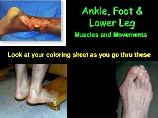
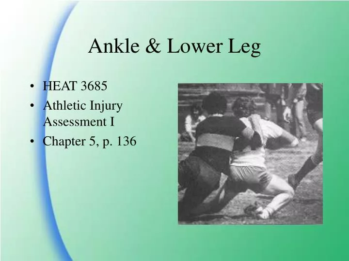
Ankle & Lower Leg. HEAT 3685 Athletic Injury Assessment I Chapter 5, p. 136. Bones: tibia fibula talus Ligaments: ATFL PTFL CFL Deltoid. Clinical Anatomy-- p.136. Interosseous membrane Muscles: peroneals anterior tibialis posterior tibialis triceps surae Bursae.

E N D
Ankle & Lower Leg • HEAT 3685 • Athletic Injury Assessment I • Chapter 5, p. 136
Bones: tibia fibula talus Ligaments: ATFL PTFL CFL Deltoid Clinical Anatomy--p.136 • Interosseous membrane • Muscles: • peroneals • anterior tibialis • posterior tibialis • triceps surae • Bursae
History--p. 145 • Onset of pain-- • acute/gradual/chronic/ re-injury • PMH? (tx/rehab) • Mechanism: • INV • EV • DF • PF • Combination • Location of pain-- • Table 5-2, p.147 • medial/lateral: • probable sprain • avulsion fx • stress fx • muscle strain • Change in activity? • Position/requirements/ duration/frequency/ surface
Observation/Inspection--p.147Fig. 5-15, p. 149 • WB status (antalgic?) • Bilateral comparison • malleoli • sinus tarsi (p.fig. 5-16, p149) • triceps surae • Achilles tendon • Inflammation • Swelling • Deformity
Lateral ligaments (p.151) ATFL PTFL CFL Medial Ligaments Deltoid group Dorsalis pedis pulse fig. 5-31, p. 180 Between 1st & 2nd mets. Palpation-- p. 150-151 • Tibio-fibular Ligaments (p.152) • Anterior • Posterior
AROM: DF/PF: landmarks DF=20º PF=~50º fig. 4-24, p. 106 INV/EV: landmarks INV~20º EV~5º RROM: Box 5-3, p. 156 Functional Testing--p. 154 • Other Tests: • Toe Raise • Heel Raises • Walking/Hopping/Jumping
On-field Functional Testing • Willingness to: • Move joint • Bear weight • Contraindicated: • Obvious deformity • Be cautious when full AROM present
Ligamentous Testing--p.157 • Anterior Drawer • Posterior Drawer • Talar tilt • Kleiger test
Anterior Drawer--p.158 • Box 5-4, p. 158 • 2 methods • knee flexed/foot stabilized • Assesses laxity in ATFL • (+)= anterior movement of talus on mortise
Anterior Drawer Testing • Knee flexed/foot stabilized • Assesses laxity in ATFL • (+)= anterior movement of talus on mortise
Posterior Drawer • Similar to Anterior Drawer • Tests integrity of PTFL • (+) = Posterior movement of talus on mortise
Talar Tilt—p.159 • Inversion stress test • Box 5-5, p. 159 • Stresses CFL • Always compare bilaterally • (+) = excessive PROM in INV • Also used in x-rays (WNL: <9º) • (EV talar tilt tests the deltoid ligament.—Box 5-6, p.160)
Kleiger test—p. 161 • Box 5-7, p.161 • Syndesmosis test • External Rotation (ER) with plantar flex. (PF) • (+) Results: (2 possible) • Medial pain=deltoid sprain • Anterior pain=ant. tibiofibular sprain
Lower Leg Special Tests • Bump Test--p.170 • Squeeze test--p.166 • Thompson test--p.177 • Homan’s sign--p.181
Bump Test--p.170 • Percussion test –Box 5-9 • Ankle DF to neutral and tap calcaneus • (+) Results= • Pain proximally with distal percussion • Impression: lower leg fx. (tibia, fibula, or talus) • false positives common
Squeeze test--p.166—Box 5-8 • Compression test • Compress tibia & fibula together progressing distally • (+) Results: • Distal pain with proximal compression • Impression: lower leg fx (tibia or fibula) • Sometimes (+) with stress fx
Thompson test--p.177, Box 5-10 • Athlete is prone • Squeeze the triceps surae belly and observe passive plantarflexion • (+)=Reduced motion at ankle • Impression: torn Achilles tendon
Homan’s sign--p.181, Box 5-11 • Assesses presence of deep vein thrombosis (DVT) • Findings must agree with other symptoms • Passive DF with full knee EXT. • (+) = intense pain in calf along with other signs of inflammation • Triceps surae strain may give false (+)
Neurological Testing--p.162 • Most common neuro. Trauma: • common peroneal nerve injury • Dec. in PF, EV, DF strength • secondary to other injuries (fx, contusions, LBP) • Signs/symptoms: • Decreased strength • Paresthesia/Anesthesia • Decreased reflexes
Pathologies • Inversion ankle sprain • Eversion ankle sprain • Lower leg fractures • Stress fx • Ankle impingement • Achilles tendonitis • Subluxating Peroneal Tendons • Anterior compartment syndrome • Medial Tibial Stress Syndrome (Shin Splints)
Inversion ankle sprain--Box 5-6, p. 163 • More common than EV sprains • Mechanism: INV +/- rotation • Injured structures: ATFL/PTFL/ CFL • Symptoms: • lateral swelling/pain • hx of tight heel cord (HC) • Testing: • (+) Anterior Drawer • (+) Talar tilt • (-) Bump/Squeeze test • (-) Kleiger test • R/O fx’s in kids
Eversion ankle sprain--Box 5-8, p. 168 • Mechanism: EV +/- rotation or compression mechanism • Injured structures: Deltoid Ligament complex • Symptoms: • Medial swelling/pain • hx of tight heel cord (HC) • Testing: • (+)Kleiger test • (+) Talar tilt • (-) Bump/Squeeze test • R/O “knock-off” fx’s (p. 168)
Syndesmosis Sprains(High Ankle sprains) • 10 – 18% of cases • Involves the Ant/Post. Tib/fib. Ligaments, interosseous membrane, crural interosseous ligament, possibly deltoid lig. • MOI – excessive ER of talus with associated DF • S/S – pain at anterior/dist aspect of lower leg. Inc with DF, ER and squeeze test
Syndesmosis Sprains(High Ankle sprains) • Eval – palpate entire shaft of fib for crepitus • FX – usually in distal 1/3 but can be in proximal 1/3 (Maisonneuve FX) • TX – splint & crutch • Recovery: usually 3 – 4 weeks
Ankle Impingement Syndrome • Hx--recurrent ankle sprains • Symptoms: • Tenderness between Ant. Tib. Tendon and Med. Malleolus • chronic inflammation • Pain worsens with DF and decreases with PF • Ankle weakness in INV/EV • Anterior pain without laxity
Stress Fractures • History: • gradual onset • Usually accompanies a change in activity • c/o “burning” after activity in lower leg • Palpation: • point tenderness at site of fx • often confused with other injuries • Observation: • Swelling is minimal/absent • Altered gait/activities due to pain • Usually no discoloration/deformity • Special Tests: • (-) Bump test? • (-) Squeeze test? • (+) Tuning Fork sign • Tx: Minimum 2 wks rest
Achilles Tendinitis--p.173 • History: • Poorly vascularized area • Usual mechanism= overuse • Sometimes acute (strain/trauma) • Check shoes, gait, and technique for risk factors • Palpation: • Usually point tender at musculotendonous junction • Crepitus possible with AROM • Observation: • Localized inflammation which worsens with activity • Over pronation/supination • Special Tests: • Thompson test painful • Limited AROM in DF/PF • If untreated, may lead to HC rupture
Achilles Tendinitis--p.173 Treatment: Eliminate cause(s) Temporary heel lift Gentle stretches (2) Arch supports Taping Modalities & Medications
Box 5-14 May be primary or secondary injury Subluxation may be seen, felt, or heard Easily palpated with AROM and RROM Fig. 5-30: Biomechanical changes possible May require surgical correction to prevent further injuries Subluxating Peroneal Tendon—p. 178
Anterior compartment syndromep. 179 • History: • Acute or chronic onset • Traumatic or overuse onset • C/O numbness/tingling in foot with decreased DF • Palpation: • Decreased dorsalis pedis pulse • Dec. RROM in DF • Observation: • Possible generalized swelling • Altered gait due to pain and weakness • Treatment: • Find/eliminate cause • Avoid ext. compression • Decrease int. compression • fasciotomy may be indicated
Shin Splints • Medial tibial stress syndrome • Pain with activity in distal 1/3 of tibia • Initially pain only after activity • Two primary causes: • Overuse (Muscle imbalance) • Biomechanical (Overpronation)
Medial Tibial Stress Syndrome(Shin Splints) • Caused by Overuse: • Evaluate PROM in DF and PF • Evaluate Achilles flexibility • Treatment: • Improve circulation in lower leg • Reverse muscle strength imbalance
MTSS cont. • Caused by Poor Biomechanics: • Evaluate RROM in EV and INV • Evaluate Achilles flexibility • Assess arch integrity • Treatment: • Improve circulation in lower leg • Support arch • Strengthen post. tib.