Download
1 / 77
780 likes | 1.02k Vues
Lower Leg, Ankle, and Foot Conditions. Chapter 19. Anatomy. Anatomy. Anatomy. Anatomy (cont.). Hindfoot Calcaneus and talus Talocrural joint (ankle joint) Articulation of talus, tibia, and fibula Close-packed position—dorsiflexion Medial ligament—deltoid

E N D
Lower Leg, Ankle, and Foot Conditions Chapter 19
Anatomy (cont.) • Hindfoot • Calcaneus and talus • Talocrural joint (ankle joint) • Articulation of talus, tibia, and fibula • Close-packed position—dorsiflexion • Medial ligament—deltoid • Lateral ligament—anterior talofibular; posterior talofibular; calcaneofibular
Anatomy (cont.) • Tibiofibular joints • Superior—proximal • Inferior—distal • Interosseous membrane
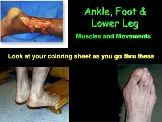
Anatomy (cont.) • Muscles • Lateral and medial view
Anatomy (cont.) • Muscles • Posterior view
Anatomy (cont.) • Nerves • Sciatic nerve • Tibial nerve • Common peroneal nerve — deep and superficial peroneal nerves • Femoral — saphenous
Anatomy (cont.) • Blood supply • Femoral artery • Popliteal • Anterior and posterior tibial • Anterior tibial • Dorsal pedal
Kinematics (cont.) • Motions • Ankle— dorsiflexion and plantarflexion • Subtalar joint • Inversion and eversion • Pronation-combination of dorsiflexion, eversion and abduction • Supination-combination of plantar flexion,inversion, and adduction
Lower Leg Contusions • Gastrocnemius contusion • S&S • Immediate pain and weakness • Rapid hemorrhage and muscle spasm → palpable mass • Management: cold with gentle stretch • Tibial contusion (shin bruise) • Vulnerable lack of padding • Minor injury—caution: repeated blows → damage periosteum • Key: prevention
Lower Leg Contusions (cont.) • Acute compartment syndrome • Lower leg includes 4 nonyielding compartments • Mechanism: direct blow anterolateral aspect of the tibia • Consequence: rapid ↑ in tissue pressure → neurovascular compromise
Lower Leg Contusions (cont.) • S&S • History of trauma • Increasingly severe pain—out of proportion to situation • Firm and tight skin over anterior shin • Loss of sensation between 1st and 2nd toes on dorsum of foot • Diminished pulse—dorsalis pedis artery • Functional abnormalities within 30 minutes • Management: cold; no compression or elevation; immediate physician referral • Irreversible damage can occur within 12–24 hours
Ankle Sprains • Inversion ankle sprain • Mechanism: plantarflexion and inversion • Predisposing factors • Lateral malleolus projects farther downward • Weakness in peroneals • ↓ ROM in Achilles tendon
Ankle Sprains (cont.) • Eversion ankle sprain • Mechanism: excessive dorsiflexion and eversion • Deltoid ligament • Potential • Lateral malleolus fracture; bimalleolar fracture • Tear of anterior tibiofibular ligament and interosseous membrane • Predisposing factors • Excessive pronation • Hypomobile foot
Ankle Sprains (cont.) • S&S (eversion sprain) • Mild to moderate injuries • Often unable to recall the mechanism • Some initial pain at time of injury, but often subsides and individual continues to play
Ankle Sprains (cont.) • Swelling • May not be as evident as a lateral sprain • Between posterior aspect of lateral malleolus and Achilles tendon • Point tenderness in involved ligaments • Severe injuries • PROM pain-free in all motions except dorsiflexion
Ankle Sprains (cont.) • Syndesmosis sprain • Spreading of space at distal tibiofibular joint • Mechanism: dorsiflexion and external rotation • Common: anterior inferior tibiofibular ligament • Assessment based on: • External rotation test • Squeeze test • Syndesmosis ligament palpation • Passive dorsiflexion test
Ankle Sprains (cont.) • Management of ankle sprains • Standard acute • Assessment for additional damage (e.g., fracture) • Use of appropriate immobilization • Moderate/severe—physician referral
Ankle Sprains (cont.) • Subtalar dislocation • Results from a fall from a height (as in basketball or volleyball); foot lands in inversion • disrupts interosseous talocalcaneal and talonavicular ligaments
Ankle Sprains (cont.) • S&S • Extreme pain and total loss of function is present • Gross deformity may not be clearly visible • Foot may appear pale and feel cold to the touch • Individual may show signs of shock • Concern: potential for peroneal tendon entrapment and neurovascular damage • Management: medical emergency; activate EMS; monitor neurovascular function
Strains of Foot and Lower Leg • Tendinitis • Common sites • Achilles tendon just proximal to insertion on calcaneus • Tibialis posterior just behind medial malleolus • Tibialis anterior on dorsum of foot just under extensor retinaculum • Peroneal tendons just behind lateral malleolus and at distal attachment on base of 5th metatarsal
Strains of Foot and Lower Leg (cont.) • Predisposing factors • Training errors • Direct trauma • Infection from a penetrating wound into tendon • Abnormal foot mechanics producing friction between shoe, tendon, and bony structure • Poor footwear that is not properly fitted to foot
Strains of Foot and Lower Leg (cont.) • S&S (tendinitis) • History of morning stiffness • Localized tenderness over tendon • Swelling or thickness in tendon and peritendon tissues • Pain with passive stretching and with active and resisted motion • Management • Cryotherapy • Address any mechanical problems
Strains of Foot and Lower Leg (cont.) • Peroneal tendon strain • Mechanism • Strong push-off a slightly pronated foot • Forceful passive dorsiflexion • Direct blow—posterior lateral malleolus • Retinaculum tears, tendons slip forward over lateral malleolus; simultaneous reduction
Strains of Foot and Lower Leg (cont.) • S&S • Cracking sensation followed by intense pain and inability to walk • Swelling and point tenderness in posterior superior lateral malleolus • Extreme discomfort or apprehension during attempted eversion against resistance • Chronic—complains of “giving way” with little discomfort
Strains of Foot and Lower Leg (cont.) • Tibialis posterior tendon strain • S&S • Pain, mild swelling • Weakness in plantarflexion and inversion • Aids in supporting the MLA • Could lead to collapse of midfoot; hyperpronation may be visible
Strains of Foot and Lower Leg (cont.) • Gastrocnemius strain • Medial head or musculotendinous junction • Mechanism • Forced dorsiflexion while knee is extended • Forced knee extension while foot is dorsiflexed • Muscular fatigue with fluid–electrolyte depletion and cramping • S&S • Immediate pain, swelling, loss of function • Management: standard acute; gentle stretching; heel lifts
Strains of Foot and Lower Leg (cont.) • Achilles tendinitis • Risk factors • Tight heel cords • Foot malalignment deformities • Recent change in shoes or running surface • Sudden increase in workload or change in exercise environment
Strains of Foot and Lower Leg (cont.) • Acute S&S • Aching or burning pain in posterior heel, ↑ with passive dorsiflexion and resisted plantarflexion • Point tenderness and crepitus at bony insertion • Local nodules • Chronic S&S • Pain worse after exercise • Thickened tendon • Tightness in gastrocnemius–soleus • Management: cryotherapy; NSAIDs; activity modification
Strains of Foot and Lower Leg (cont.) • Achilles tendon rupture • Mechanism: push-off of forefoot while knee is extending • More common in athletes over age 30
Strains of Foot and Lower Leg (cont.) • S&S • “Pop” • Inability to stand on toes • Visible defect • Excessive passive dorsiflexion • + Thompson’s test • Management • Compression wrap and splint; immediate physician referral
Overuse Conditions (cont.) • Medial tibial stress syndrome • Periostitis along posteromedial tibial border (distal third) • Believed to be related to periostitis of the soleus insertion along the posterior medial tibial border • Excessive pronation causes an eccentric contraction of soleus → periostitis • Other contributing factors • Recent changes in running distance, speed, footwear, or running surface
Overuse Conditions (cont.) • S&S (MTSS) • Dull pain begins at any point in the workout; occasionally sharp and penetrating • Pain along posteromedial border of tibia in distal third • Pain is relieved with rest, but may recur hours after activity stops
Overuse Conditions (cont.) • Secondary to mechanical abnormalities: • Increased Achilles tendon angle • Greater Achilles tendon angle between heel strike and maximal pronation • Greater passive subtalar motion in inversion and eversion • ↑ pain with active plantarflexion • Management: rest!!! cryotherapy; NSAIDs; refer to Application Strategy 19.5
Overuse Conditions (cont.) • Exertional compartment syndrome • Characterized by exercise-induced pain and swelling that is relieved by rest • Compartments most frequently affected—anterior (50%–60%) • Usually seen in well-conditioned individuals younger than 40
Overuse Conditions (cont.) • S&S • Aching leg pain and sense of fullness over involved compartment • Often affects both legs • Symptoms relieved with cessation of exercise • Activity-related pain begins at a predictable time • Anterior compartment—mild foot drop; paresthesia on dorsum of the foot • Perform evaluation after exercise strenuous enough to reproduce symptoms • Management: assessing contributing factors
Venous Disorders • Deep vein thrombosis (DVT) • Partial or complete blockage of a vein due to accumulated blood products that form a clot • Common—deep calf veins • Embolism • Obstruction or occlusion of a vessel by bacteria or other foreign body
Venous Disorders (cont.) • DVT is typically asymptomatic and may not become apparent until a pulmonary embolism occurs • Most reliable signs • Paresthesia in the area • Chronic swelling and edema in the involved extremity, engorged veins • Ecchymosis formation with a blue hue • + Homan’s sign • Management: immediate physician referral
Neurologic Conditions (cont.) • Tarsal tunnel syndrome • Posterior tibial nerve (or branch) constricted beneath fibrous roof of foot flexor retinaculum • Often linked to excessive pronation or excessive valgus deformity • S&S • Pain at medial malleolus radiating into sole and heel • Paresthesia, dysesthesia, or hyperesthesia in nerve distribution • + Tinel’s sign • Management: rest; NSAIDs; orthoses; gradual return to activity
Foot and Lower Leg Fractures • Repetitive microtraumas → apophyseal or stress fractures • Tensile forces associated with severe ankle sprains → avulsion fractures of 5th metatarsal • Severe twisting → displaced and undisplaced fractures in foot, ankle, or lower leg
Foot and Lower Leg Fractures (cont.) • Stress fractures • Often seen in running and jumping, especially after significant ↑ training mileage; change in surface, intensity, or shoe type • Common sites • 2nd metatarsal • Sesamoid bones • Navicular • Calcaneus • Tibia and fibula
Foot and Lower Leg Fractures (cont.) • S&S • Pain begins insidiously; ↑ with activity and ↓ with rest • Pain usually limited to fracture site • Pain with percussion, tuning fork, or ultrasound • Management: standard acute; physician referral
Foot and Lower Leg Fractures (cont.) • Avulsion fractures • Eversion sprain—deltoid ligament avulses portion of distal medial malleolus • Inversion sprain—plantar aponeurosis or peroneus brevis tendon avulses base of 5th metatarsal (type II) • Jones fracture • Type I transverse fracture into the proximal shaft of 5th metatarsal at junction of diaphysis and metaphysis • Often overlooked in conjunction with a severe ankle sprain • Complications: nonunions and delayed unions are common • Management: standard acute; physician referral