Download
1 / 48
480 likes | 734 Vues
Injuries to the Lower Leg, Ankle, and Foot. Injuries to the Lower Leg, Ankle, and Foot…. For an athlete to move well, there must be excellent functioning of the lower leg, ankle, and foot The foot must provide a stable base of support and as the same time be flexible and extremely mobile

E N D
Injuries to the Lower Leg, Ankle, and Foot… • For an athlete to move well, there must be excellent functioning of the lower leg, ankle, and foot • The foot must provide a stable base of support and as the same time be flexible and extremely mobile • This chapter discusses the skeletal and muscular anatomy of the foot and lower leg • We will discuss: • Ligaments of the ankle, compartments of the lower leg, muscular actions of each compartment • Fractures as well as common sprains of ankle ligaments
Injuries to the Lower Leg, Ankle, and Foot… • Treatment of ankle sprains and control of possible future sprains • Recognition, care, and treatment of tendon injuries along with compartment problems • Treatment and care of athletes with shin splints and considers ways to enhance the performance of these athletes • Discuss foot disorders such as plantar fasciitis, heel spurs, Morton's neuroma, arch problems, bunions, blisters and calluses, providing guidelines for recognition, first aid treatment, and long term care • And FINALLY ANKLE TAPING
Anatomy Review • The lower leg, ankle, and foot work together to provide a stable base of support and a dynamic system of movement • The skeleton of the lower leg consist of the tibia and fibula
Anatomy Review • Tibia is the larger and stronger of the two (commonly called the shin bone) • Supports 98% of body wgt • Acts as an attachment for various muscles and helps to provide a mechanical advantage for some of them
Anatomy Review • Normal foot contains 26 bones that are interconnected and supported by numerous ligaments • Many joints within the foot also assist with support and movement
Anatomy Review • The ankle joint (talocrural joint) is where the tibia, fibula, and talus join • Provides mainly plantar flexion and dorsiflexion of the foot • Subtalar joint is the articulation of the talus and the calcaneus • Responsible for inversion and eversion of the foot • Both joints are synovial, which means they are surrounded by a capsule and supported by ligaments
Anatomy Review • The ankle joint is supported on the medial side by the large and strong deltoid ligament • On the lateral side, the joint is supported by the anterior talofibular, the posterior talofibular, and the calcanefibular ligaments
Anatomy Review • These ligaments are not as large or strong as the deltoid ligament • Additional lateral stability for the ankle joint is provided by the length of the fibula on the lateral side of the ankle • The ankle joint is strongest when placed in dorsiflexion • The talus fits much tighter between the tibia and fibula in this position • Joint is weakest when placed in plantar flexion
Anatomy Review • Joints, ligaments, and muscles help to create and maintain the two basic arches in the foot • Longitudinal arch has medial lateral divisions • Transverse arch runs from side to side • These arches assist the foot as shock absorbers; also provide propulsion off surfaces during movement
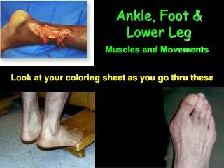
Anatomy Review • Muscles are divided into anterior (front), posterior (back), and lateral (side) compartments • Muscles of the anterior compartment essentially produce dorsiflexion and extension of the toes • Tibialis anterior, extensor digitorumlongus, extensor hallucislongus, and peroneustertius • Very compact area with little room for any extra tissue or fluid
Anatomy Review • Posterior compartment mainly functions to produce plantar flexion of the foot • Referred to as the calf muscles • Is divided into two compartments, superficial section and deep section • Superficial section • Gastrochnemius, soleus, and plantar muscles • Gastrochnemius and soleus attach on the calcaneus via the achilles tendon • Plantars muscle is small and insignificant in action
Anatomy Review • Deep section of this compartment houses the tibialis posterior, flexor digitorumlongus, flexor hallucislongus, and popliteus muscles • Besides the popliteus these muscles course behind the medial mallelous of the tibia and along the bottom of the foot • They help with the plantar flexion as well as flexion of the toes • The popliteus muscle is important in knee flexion
Anatomy Review • Lateral compartment of the lower leg contains the peroneuslongus and peroneusbrevis muscles • Mainly evertors (to turn the foot outward) of the foot but do assist with some plantar flexion • Both of these muscles course behind the lateral mallelous of the fibula • Peroneuslongus courses under the lateral side of the foot and runs across the bottom to the first metatarsal and cuneiform bones • The peroneusbrevis attaches at the base of the 5th metatarsal and is subject to avulsion (forcible tearing away or separation)
Anatomy Review • Included is also the peroneal nerve, a superficial nerve that is susceptible to injury • The posterior tibial artery supplies blood to the peroneal muscles because there is no major artery in the lateral compartment
Common Sports Injuries • Many injuries occur to the lower leg, ankle, and foot • Some can be classified as traumatic, and others are chronic in nature • Traumatic injuries typically involve skeletal structures • Chronic injuries usually involve damage to soft tissues
Skeletal Injuries…Fractures • Direct trauma through contact causes most fractures to the lower leg • Magnitude of contact necessary to fracture a bone such as the tibia or fibula can vary • A fracture can be caused by being kicked by an opponent in a soccer match or by having a 300 pound lineman land on a leg • http://www.youtube.com/watch?v=I-iEOoM1N-w
Skeletal Injuries…Fractures • Fractures to the foot can also occur from trauma • However, violent trauma is not always required in fractures of the bones of the leg and foot • Stress fractures can occur from overuse or microtrauma (microscopic lesion/injury)
Skeletal Injuries…fractures • In running, for example, each time the foot strikes the ground it produces a small amount of trauma to the bone • This trauma damages a few bone cells, which the body must repair as quickly as possible • When the body cannot maintain the repair process and keep up with repeated microtrauma to a specific bone, a stress fracture results • Additionally, an avulsion fracture of the 5th metatarsal can occur in association with a lateral ankle sprain • Therefore the possibility of such a fracture should be examined when an athlete sprains his/her ankle
Skeletal Injuries…fractures • S&S • Swelling and/or deformity at the location of the trauma • Discoloration at the site of the trauma • Possible broken bone end projecting through the skin • Athlete reports that a snap or a pop was heard or felt • The athlete may not be able to bear weight on the affected extremity • In the case of a stress fracture or a growth plate fracture that did not result from a traumatic event, the athlete complains of extreme point tenderness and pain at the site of suspected injury
Skeletal Injuries…fractures • TX: • Watch and treat for shock if necessary • Apply sterile dressings to any related wounds (ex open fx) • Carefully immobilize the foot and leg using a splint • Arrange for transport to a medical facility • In the event that bones are fractured, apply a cast • Athlete will be immobilized for a specified time
Skeletal Injuries…fractures • When the fracture has healed properly, the physician will release the athlete for rehabilitation, practice, and competition in that order • Participation while a fracture is healing is NOT recommended because it may slow the healing process • There is a possibility of nonunion of a fracture, especially in the 5thmetartasal of the foot, as a result of a diminished blood supply
Soft-Tissue Injuries…ankle injuries • One of the most common sports injuries to the lower leg and ankle is a sprained ankle • Are abnormal stresses placed on ligamentous structures and cause various levels of damage • Sprains can occur to the lateral or medial ligaments of the ankle depending on which direction the foot moves when abnormal stress is placed on the ligaments and the foot rolls to one side
Soft-Tissue Injuries…ankle injuries • The noncontractile structures on the lateral aspect of the ankle are most susceptible to injury • The formation of the bones of the ankle helps to stabilize it; the fibula extends inferiorly, approximating the lateral talus completely • The ligaments on the lateral side, the anterior talofibular, the posterior talofibular, and the calcaneofibular ligaments are not as large or strong as the deltoid ligament on the medial side of the ankle joint
Soft-Tissue Injuries…ankle injuries • It is estimated that 80% to 85% of ankle sprains occur to the lateral ligaments (Ryan et al., 1986) • An interesting note is that authors are suggesting that serious ankle sprains in the adolescent athlete are unusual because the ligaments are typically stronger than the bones (Omey & Micheli, 1999)
Soft-Tissue Injuries…ankle injuries • Can occur in virtually any sport and can limit the abilities of the athlete in performance until resolution of the injury is complete • As the severity of the ankle sprain increases, so does the instability of the ankle • It is generally accepted that an eversion (move outward) ankle sprain is more severe, with greater instability, and should be cared for more conservatively (Ryan et al., 1986) • However, an inversion (move inward) ankle sprain is more common, with the lateral ligaments being involved in 80% to 85% of all ankle sprains
Soft-Tissue Injuries…ankle injuries • S&S of a lateral ankle sprain • 1st degree sprain • pain, mild disability, point tenderness, little laxity, little or no swelling • 2nd degree sprain • Pain, mild-moderate disability, point tenderness, loss of function, some laxity (abnormal movement), swelling (moderate to severe)
Soft-Tissue Injuries…ankle injuries • 3rd degree sprain • Pain and severe disability, point tenderness, loss of function, laxity (abnormal movement), swelling, (moderate to severe)
Soft-Tissue Injuries…ankle injuries • TX: • Immediately apply ice, compression, and elevation • A horse-shoe or doughnut shaped pad kept in place by an elastic bandage aids at this stage in the compression and reduction of fluid • Have the athlete rest and use crutches to ambulate with a 3 or 4 point gait if a 2nd or 3rd degree sprain has occurred • If there is any hesitation about the severity, splint and refer for further eval
Soft-Tissue Injuries…ankle injuries • It is important to recognize the possibility of a tibiofibular (tib/fib) syndesmosis sprain in conjunction with or masquerading as a lateral ankle sprain • Too often a syndesmosis sprain is treated as a lateral ankle sprain, which is inappropriate and will not allow the athlete to progress in the healing process
Soft-Tissue Injuries…ankle injuries • It is important to know that there is a significant difference in the etiology of the injury • With the lateral ankle sprain, there is an inversion mechanism, which includes supination • In the tib-fib syndesmosis sprain, the mechanism is one of dorsiflexion followed by axial loading of the lower leg, with external rotation of the foot and internal rotation of the lower leg • Typically, athletes have their foot planted firmly with the foot in external rotation, and the lower leg twist medially, forcing the talus into the ankle mortise • The axial load forces the tibia and fibula to separate slightly and sprain the syndesmosis
Soft-Tissue Injuries…ankle injuries • S&S of a tib/fib syndesmosis sprain • Mechanism of injury is different from a lateral ankle sprain; ankle dorsiflexion and foot external rotation are combined with internal rotation of the lower leg • Typical ankle sprain test may be positive but the athlete will c/o a great deal of pain and point tenderness in the area of the tib/fib syndesmosis
Soft-Tissue Injuries…ankle injuries • Performing the “squeeze” test (squeezing the tibia and fibula together superior to the syndesmosis); elicits pain in the syndesmosis area • TX • Immediately apply ice, compression, and elevation • A horse-shoe or doughnut shaped pad kept in place by an elastic bandage aids • Have the athlete rest and use crutches to ambulate for the first 72 hours, followed by use of a walking boot for a minimum of 3 days and preferably for 7 days following the initial injury • If there is any question refer for further evaluation
Soft-Tissue Injuries…ankle injuries • It is recognized that either taping or bracing can reduce the number of ankle sprains (Verhagen, van Mechelen, & de Vente, 2000) • Some prefer to use the standard ankle-taping procedure as a prophylactic tx for ankles with no HX of an injury • Others choose to augment the taping procedure to prevent future ankle sprains if one has occurred before • In published research studies, ankle taping as been demonstrated to help with the neuromuscular response of the muscles and to provide stability if done in a specific manner • Both contribute to reduction of ankle sprains
Soft-Tissue Injuries…ankle injuries • Most researchers agree that the best known method of ankle support, the prophylactic adhesive-taping procedure, supports the ankle for only a short period of time after exercise begins (Frankeny et al., 1993) • Therefore, some researchers now maintain that bracing is better than taping for the prevention of ankle injuries, owing to the reduction in ROM, either at excessive points or within normal ranges (Cordova, Ingersoll, & LeBlanc, 2000) • The combination of high-top shoes and taping or bracing can be helpful to athletes in reducing the number of ankle sprains they experience
Soft-Tissue Injuries…ankle injuries • Proprioception and the ankle is a very intense area of study • Proper ankle proprioception is a critical element in reducing chronic ankle instability (Hintermann, 1999) • Also be important part of both the preventative and rehabilitative aspects of ankle functioning (Hertel, 2000)
Soft-Tissue Injuries…ankle injuries • Whatever the choice of the coach or athlete, many factors must be considered in preventing ankle sprains • These include: • Type of activity, compliance of the athlete in wearing braces or prophylactic taping, cost to the school or athlete, effectiveness of the brace as reported in research studies • There are some consequences of using adhesive tape, including: • Blisters, tape cuts, and loss of circulation
Tendon-related Injuries • The achilles tendon is commonly injured by long distance runners, basketball players, and tennis players • The onset of tendinitis may be slow among runners, but much more rapid among basketball or tennis players • Great many of short-burst movements requiring jumping or rapid motion from side to side
Tendon-related injuries • Some controversy exsist about the actual injury that constitutes Achilles tendinitis • The Achilles tendon itself, which attaches the gastrocnemius and soleus muscles to the calcaneus, can become inflamed • However, either tendon sheath or the subcutaneous bursa dorsal to the tendon can become inflammed
Tendon-related injuries • Most agree that athletes who dramatically increase their running distance or workout times and who do so running on hard, uneven, or uphill surfaces are prone to Achilles tendinitis (Omey & Micheli, 1999) • It is estimated that 11% of runners and up to 52% of former elite runners experience an Achilles tendinopathy (tiny tears (microtears) in the tissue in and around the tendon caused by overuse) • http://www.youtube.com/watch?v=F2e6LmQsJps
Tendon-related injuries • Superficially, Achilles tendinitis can produce an increased temperature in the immediate area; moreover, the tendon is painful on touch and movement and appears thickened • This pain associated with this condition is localized to a small area of the tendon and typically intensifies when movement is initiated after rest • Can be seen over an extended period of time (days to weeks) • Or over a shorter period of time (days)
Tendon-related injuries • TX for chronic Achilles tendinitis: • Immediate rest until the swelling subsids • NSAIDS, small heel lift assist the reduction of swelling and the return to practice and competition • Stretching has also been shown to be beneficial to athletes with Achilles Tendinitis (Taylor et al., 1990) • Controlled stretching on a slant board or against a wall each day will aid in a return to participation • Additionally, if an athlete must exercise or run, it is advised this be done in a controlled environment (swimming pool)
Tendon-related injuries • Controlled gradual stressing exercises using the eccentric contraction of the Achilles assist the athlete in returning to activity • An athletes activity level and type of exercise must be closely monitored during the healing phase • Without the proper amount of rest, the body has a hard time repairing injury, thereby increasing the amount of time the athlete experiences difficulty with the condition
Tendon-related injuries • Explosive jumping or direct trauma from some type of impact can cause traumatic injuries to the Achilles tendon by tearing or rupturing the tendon • Can occur in many different sports
Tendon-related injuries • S&S • Swelling and deformity at the site of injury • Reports a pop or snap associated with the injury • Pain in the lower leg, which may range from mild to extreme • Loss of function, mainly in plantar flexion • TX: • Immediately apply rice and compression to the area • Immobilize the foot by an air cast or splint • Arrange for transportation to the nearest medical facility
Tendon-related issues • During the acute phase of the healing process, minimize dorsiflexion and eliminate forced dorsiflexion • This movement can produce more damage and inflammation to the area • The long term effects of a ruptured Achilles tendon depend on the severity or completeness of the rupture • If surgery is necessary, the athlete will most likely be out of commission for the rest of the season • The athlete will need to be careful and aware of the value of stretching and warming up