Download

1 / 32
320 likes | 538 Vues
Aortorenal & Selective Renal Arteriography. performed by percutaneous needle puncture Catheterization of common femoral arteries Indications for renal arteriography include Suspected renal artery stenosis (renovascular hypertension) Vascular malformations

E N D
Aortorenal & Selective Renal Arteriography • performed by • percutaneous needle puncture • Catheterization of common femoral arteries • Indications for renal arteriography include • Suspected renal artery stenosis (renovascular hypertension) • Vascular malformations • tumor embolization to minimize surgical • blood loss or treat bleeding tumors • Trauma
Diagnostic renal angiography to demonstrate renal vascular anatomy • Complications from conventional catheter angiography include • bleeding at the puncture site • contrast allergy or nephrotoxicity • renal or distal emboli.
Normal abdominal aortogram Bilateral renal artery stenoses
3D coronal CT angiography image demonstrates an inferior accessory left renal artery (posterior view) The left accessory renal artery origin (*) is better demonstrated rotating the model in the axial plane
Inferior Venacavography & SelectiveVenography • Risks of bleeding and emboli present in arterial studies are virtually eliminated • Venography • rarely used today since theinformation can be obtained at cross-sectional imaging • (CT or MRI) in almost all cases • Adrenal and renal venography • performed occasionally in the setting of venous sampling to localize hormone secretion in patients with indeterminate noninvasive imaging studies
Normal inferior vena cava (C) Unusual retrograde filling of morphologically normal renal veins (arrows) from antegrade injection into the inferior vena cava B = retained contrast material in the cecum from previous barium enema examination Inferior vena cava obstruction
Double inferior vena cava (R, L). Persistent left supracardinal vein anomaly Example of duplicated IVC on IV contrast enhanced axia
Angiography: renal venography. Normal left renal vein Tumor thrombus
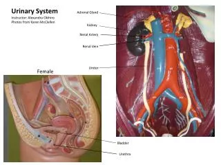
Miscellaneous Urologic Angiography • Although angiography has little or no value in examination of the ureter, bladder, adrenals, and prostate, angiograms of these structures may be indicated in particular clinical situations, in which case the studies are usually “tailored” to the clinical problem • Rarely used • Corpus cavernosograms are made by direct injection of suitable contrast material into the corpora cavernosa of the penis • useful in examining for Peyronie’s disease, • impotence, priapism, and traumatic penile lesions • but these also are not commonly performed
SONOGRAPHY • Ultrasound waves for imaging are generated by transducers, devices that convert electrical energy to sound energy and vice versa • Special piezoelectric crystals that emit ultrasonic waves when they are deformed by an electrical voltage and, conversely, generate an electrical potential when struck by reflected sound waves • act as both sonic transmitters and detectors • Ultrasound images are reflection images formed when part of the sound that was emitted by the transducer bounces back from tissue interfaces to the transducer • A more sensitive method of detecting flow, called • Power mode Doppler
Clinical Uses • evaluation of the kidney, urinary bladder, prostate, testis, and penis • assessing renal size and growth • triaging patients with renal failure • detection and characterization of renal masses • provides an effective method of distinguishing benign cortical cysts from potentially malignant solid renal lesions • used to follow up mildly complicated cysts detected on CT
Doppler sonography • is useful for the evaluation of renal vessels, vascularity of renal masses, and complications following renal transplant • detect • renal vein thrombosis • renal artery stenosis • ureteral obstruction prior to the development of hydronephrosis • arteriovenous fistulas • pseudoaneurysms
COMPUTED TOMOGRAPHY SCANNING • thin, collimated beam of x-rays is passed through the patient and captured by an array of solid-state or gas detectors • Spiral (or helical) CT uses a slip-ring gantry that rotates continuously while the patient moves constantly through the collimated x-ray beam • ability to image during specific phases of contrast bolus enhancement • allows improved image reformations • Multidetector, or multislice, helical CT scanners have an array of multiple rows of detectors in a helical scanner such that multiple scan images can be acquired per gantry rotation, and as a by-product thinner sections and higher resolution achieved
Clinical Uses • used in the evaluation of: • acute flank pain • Hematuria • renal infection (search for abscess) • renal trauma • characterization and staging of renal neoplasm • evaluation of renal anatomy and pathology • Contrast media is usually administered
CT densitometry. Thin section CT of incidental right adrenal mass Normal adrenal glands Bilateral adrenal lymphoma Left adrenal carcinoma Coronal oblique reformatted image from the same patient shows the mass (arrow) to be inseparable from the inferior left adrenal limb Axial CT image reveals a predominant solid mass (arrow) abutting the left kidney, with areas of cystic change
CT scans: kidneys Simple renal cyst New Hydronephrosis
MAGNETIC RESONANCE IMAGING • Widely used in assesing/ ealuating renal masses and its potential for non invasive vascular imaging
Clinical Uses • Demonstration of congenital anomalies • diagnosis of renal vein thrombosis • diagnosis and staging of renal cell carcinoma • MR angiography • useful in evaluating renal transplant vessels • renal vein tumor or thrombosis • renal artery stenosis
primarily to stage bladder tumors and to differentiate between benign bladder wall hypertrophy and infiltrating malignant neoplasm • principally used to stage patients with prostate cancer
Indication • Evaluation • Renal masses • Renal vasculature • Prostate Cancer • Adrenal Masses
Contraindication • Presence of ferromagnetic intracranial vascular clips, cardiac pacemaker, and certain prosthetic heart valves • Severe claustrophobia and the patient’s inability to lie still 30-45 minutes of imaging(relative contraindication)
COMPARISON OF IMAGING METHODS • As new imaging methods have been developed, changes have occurred in patterns of use for each type of imagin • increased familiarity with and confidence in sonography and CT scanning have resulted in a decrease in the use of some long-established conventional uroradiologic studies such as EU.
Several factors are involved in these changes: • (1) the increased effectiveness of newer imaging methods over older ones for some aspects of urodiagnosis • (2) the availability of equipment, trained technical personnel to operate it, and physicians to interpret the results • (3) increased awareness of the hazards of ionizing radiation • (4) the desire to use noninvasive examinations if possible
Comparison of imaging methods in the evaluation of renal cell carcinoma Computed tomography (CT) scan Angiogram MRI scans MRI scans
Comparison of imaging methods Right femoral arteriogram Excretory urogram CT scan CT scan