Download

1 / 71
830 likes | 1.47k Vues
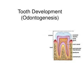
Development of Tooth and Supporting Tissues Raj Gopalakrishnan B.D.S., Ph.D. Oral and Maxillofacial Pathology Dept. of Diagnostic and Biological Sciences University of Minnesota School of Dentistry. Tooth and Associated Structures. Tooth Development. Bud Stage Cap Stage Bell Stage

E N D
Development of Tooth and Supporting Tissues Raj Gopalakrishnan B.D.S., Ph.D. Oral and Maxillofacial Pathology Dept. of Diagnostic and Biological Sciences University of Minnesota School of Dentistry
Tooth Development • Bud Stage • Cap Stage • Bell Stage • D and E. Dentinogenesis and • amelogenesis • Crown formation • Root Formation and • eruption • H. Function Essentials of Oral Histology and Embryology, Ed: James Avery, 2nd edition. 2000.
Initiation of Tooth Development The initiation of tooth development begins at 37 days of development with formation of a continuous horseshoe-band of thickened epithelium in the location of upper and lower jaws – Primary Epithelial Band • Each band of epithelium will give • rise to 2 sub divisions: • Dental lamina and • Vestibular lamina Figure from Ten Cate’s Oral Histology, Ed., Antonio Nanci, 6th edition
Maxillary Process Stomodeum Dental lamina Mandibular process Developing Tongue http://www.usc.edu/hsc/dental/ohisto/
Dental Lamina • Dental lamina appears as a thickening • of the oral epithelium adjacent to • condensation of ectomesenchyme • 20 areas of enlargement or knobs • appear, which will form tooth buds • for the 20 primary teeth • Not all will appear at the same time. • The first to develop are those of the • anterior mandible region • At this early stage the tooth buds • have already determined their crown • morphology • Successional lamina: lamina from • which permanent teeth develop • The dental lamina begins to function • at 6th prenatal week and continues to • 15th year of birth (3rd molar) Primary epithelial band Ectomesenchyme Figures from: http://www.usc.edu/hsc/dental/ohisto/
Vestibular Lamina Figure from Ten Cate’s Oral Histology, Ed., Antonio Nanci, 6th edition
Tooth development is a continuous process, however can be divided into 3 stages: 1. Bud Stage 2. Cap Stage 3. Bell Stage
1. Bud Stage Intramembranous ossification Meckel’s cartilage • Bud stage is characterized by rounded, localized growth of • epithelium surrounded by proliferating mesenchymal cells, • which are packed closely beneath and around the epithelial buds http://www.usc.edu/hsc/dental/ohisto/
1. Bud Stage In the bud stage, the enamel organ consists of peripherally located low columnar cells and centrally located polygonal cells http://www.usc.edu/hsc/dental/ohisto/
2. Cap Stage Vestibular lamina http://www.usc.edu/hsc/dental/ohisto/
2. Cap Stage http://www.usc.edu/hsc/dental/ohisto/ Enamel Organ Dental Papilla Condensation of the ectomesenchyme immediately subjacent to the tooth bud caused by lack of extracellular matrix secretion by the cells thus preventing separation. Histodifferentiation begins at the end of cap stage. Epithelial outgrowth called Enamel Organ because it will eventually form the enamel Dental Papilla: Ball of condensed ectomesenchymal cells (it will form dentin and pulp). The peripheral cells adjacent to the inner dental epithelium will enlarge and later differentiate into odontoblasts
2. Cap Stage Enamel organ Enamel knot Dental papilla http://www.usc.edu/hsc/dental/ohisto/ Dental follicle or sac Dental follicle or dental sac is the condensed ectomesenchymal tissue surrounding the enamel organ and dental papilla. This gives rise to cementum and the periodontal ligament (support structures for tooth)
2. Cap Stage Lateral lamina Lateral Lamina: extension from the dental lamina that is connected to the enamel organ Enamel niche: It is an artifact produced during sectioning of the tissue. It occurs because the enamel organ is a sheet of proliferating cells rather than a single strand and contains a concavity filled with ectomesenchyme We can also see that the inner and the outer dental epithelium are being organized
Enamel Knot: Densely packed accumulation of cells projecting from the inner enamel epithelium into dental papilla. Exact role not known, but currently believed to be the organizational center for cusp development. Dental organ or tooth germ is a term used to constitute the structure that has enamel organ, dental papilla and dental follicle
Enamel knots are clusters of nondividing epithelial cells visible in sections of molar cap stage Enamel knot precursor cells are first noted by expression of p21 gene expression Enamel knot and enamel cord are temporary structures that disappear before enamel formation begins. It has been speculated that the function of the enamel knot and cord may be to act as a reservoir of dividing cells for the growing enamel organ
http://www.usc.edu/hsc/dental/ohisto/ 3. Bell Stage Dental lamina Outer dental epithelium Inner dental epithelium Dental papilla Dental follicle Cervical loop • Continued growth leads to bell stage, where the enamel organ resembles a • bell with deepening of the epithelium over the dental papilla • Continuation of histodifferentiation (ameloblasts and odontoblasts are defined) • and beginning of morphodifferentiation (tooth crown assumes its final shape)
3. Bell Stage (Early) Outer dental epithelium Stellate reticulum Stratum intermedium Inner dental epithelium Dental papilla http://www.usc.edu/hsc/dental/ohisto/ Inner dental epithelium: Short columnar cells bordering the dental papilla. These will eventually become ameloblasts that will form the enamel of the tooth crown by differentiating into tall columnar cells. The cells of inner dental epithelium exert an organizing influence on the underlying mesenchymal cells in the dental papilla, which later differentiate into odontoblasts. Outer dental epithelium: Cuboidal cells that cover the enamel organ. Their function is to organize a network of capillaries that will bring nutrition to the ameloblasts. In preparation to formation of enamel, at the end of bell stage, the formerly smooth surface of the outer dental epithelium is laid in folds. Between the folds, adjacent mesenchyme of the dental sac forms papillae that contain capillary loops and thus provide nutritional supply for the intense metabolic activity of the avascular enamel organ
3. Bell Stage (Early) Outer dental epithelium Stellate reticulum Stratum intermedium Inner dental epithelium Dental papilla http://www.usc.edu/hsc/dental/ohisto/ Stellate reticulum: Star-shaped cells with processes, present between the outer and the inner dental epithelium. These cells secrete glycosaminoglycans, which attract water, thereby swelling the cells and pushing them apart. However, they still maintain contact with each other, thus becoming star-shaped. They have a cushion-like consistency that may support and protect the delicate enamel organ. It is absent in the portion that outlines the root portions. Stratum intermedium: Cell layer between the inner dental epithelium and stellate reticulum which have high alkaline phosphatase activity. They assist inner dental epithelium (ameloblasts) to form enamel.
Outer dental epithelium Stellate reticulum Stratum intermedium Inner dental epithelium Dental papilla http://www.usc.edu/hsc/dental/ohisto/ Dental Papilla: Before the inner dental epithelium begins to produce enamel, the peripheral cells of the mesenchymal dental papilla differentiate into odontoblasts under the organizing influence of the epithelium. First, they assume a cuboidal shape and then a columnar form and acquire the specific potential to produce dentin. The basement membrane that separates the enamel organ and the dental papilla just prior to dentin formation is called the “membrana preformativa”
3. Bell Stage Higher power view http://www.usc.edu/hsc/dental/ohisto/
3. Bell Stage Inner dental epithelium Outer dental epithelium Cervical loop Cervical loop: Area where the inner and the outer dental epithelium meet at the rim of the enamel organ. This point is where the cells will continue to divide until the tooth crown attains its full size and which after crown formation will give rise to the epithelium for root formation. Is also called “Zone of Reflexion”. http://www.usc.edu/hsc/dental/ohisto/
3. Bell Stage Enamel knot Enamel cord Enamel cord: Pattern of enamel knot that extends between the inner and outer dental epithelium http://www.usc.edu/hsc/dental/ohisto/
3. Bell Stage Dental lamina (and the lateral lamina) will disintegrate and loose contact with oral epithelium. Sometimes, these epithelial cells will persist when they are called “epithelial pearls” or “cell rests of Serre” Clinical significance: Cysts will develop in these (eruption cysts) and prevent eruption, or they may form odontomas (tumors) or may form supernumery teeth http://www.usc.edu/hsc/dental/ohisto/
Crown Pattern Determination Future crown patterning also occurs in the bell stage, by folding of the inner dental epithelium. Cessation of mitotic activity within the inner dental epithelium determines the shape of a tooth.
Vascular and Nerve Supply during Tooth Development Vascular Supply: Clusters of blood vessels in dental follicle and papilla Clustering of vessels in papilla coincide with position of root formation Enamel organ is avascular, however vessels seen in close association in the follicle Nerve Supply: Initially noted in the dental follicle during bud to cap stage However after start of dentinogenesis, seen in dental papilla Nerve fibers do not enter enamel organ
Clinical Correlation. Several odontogenic cysts and tumors can arise • from developing tooth structures. Two such conditions are: • Ameloblastoma – which are tumors of odontogenic epithelium that • may arise from cell rests of enamel organ or from the developing • enamel organ among other things
Ameloblastoma Enamel Organ Histology resembles enamel organ epithelium with peripheral columnar ameloblast-like cells surrounding loosely arranged stellate-reticulum-like cells
Odontogenic myxoma Developing tooth • Odontogenic Myxoma: Tumor of the jaw that arise from odontogenic • ectomesenchyme. Histologically, looks similar to mesenchymal portion • of a developing tooth (dental papilla).
Formation of Permanent Dentition Successional tooth bud http://www.usc.edu/hsc/dental/ohisto/ The tooth germs that give rise to permanent incisors, canines and premolars form as a result of further proliferative activity within the dental lamina, lingual to the deciduous tooth germ The developing permanent molars have no deciduous predecessor and their tooth germs originate from the dental lamina that extends posteriorly beneath the oral epithelium after the jaws have grown
Essentials of Oral Histology and Embryology, Ed: James Avery, 2nd edition. 2000.
A timetable to remember Entire primary dentition initiated between 6 and 8 weeks of embryonic development. Successional permanent teeth initiated between 20th week in utero and 10th month after birth Permanent molars between 20th week in utero (first molar) and 5th year of life (third molar)
The development of hard tissues will also be discussed by Dr. Sandra Myers, however You must remember the following: ·Hard tissue formation starts at the late stages of the bell stage ·Differentiation of cells into odontoblasts and ameloblasts ·Dentin is formed before enamel ·Dentin initiates the formation of enamel
Bell Stage Essentials of Oral Histology and Embryology, Ed: James Avery, 2nd edition. 2000.
Hard Tissue Formation Deposition of dental hard tissues is called “apposition” After the crown attains its final shape during cap to early bell stage, the inner dental epithelial cells stop to proliferate, except the cells at the cervical loop First layer of dentin appears at the cusp tips and progresses cervically, and the columnar cells of the inner dental epithelium become elongated and show reverse polarization, with the nuclei adjacent to stratum intermediate (ameloblasts) The boundary between the odontoblasts and inner dental epithelium defines the future dentino-enamel junction http://www.usc.edu/hsc/dental/ohisto/
For dentinogenesis and amelogenesis to take place normally, the differentiating odontoblasts and ameloblasts will receive signals form each other – “reciprocal induction” • Stages of Apposition • Elongation of inner dental epithelium • Differentiation of odontoblasts • Formation of dentin • Formation of enamel
Ameloblasts First layer of enamel Dentin Odontoblasts At the same time or soon after the first layer of dentin (mantle dentin) is formed, the inner dental epithelial cells differentiate into ameloblasts and secrete enamel proteins. These proteins further will help in the terminal differentiation of odontoblasts. The ameloblasts will then start laying down organic matrix of enamel against the newly formed dentinal surface. The enamel matrix will mineralize immediately and form the first layer of enamel. The formation of enamel is called amelogenesis. http://www.usc.edu/hsc/dental/ohisto/
Apposition At the same time when the inner dental epithelium is differentiating, the undifferentiated ectomesenchymal cells increase rapidly in size and ultimately differentiate into odontoblasts The increase in size of the papillary cells leads to elimination of the acellular zone between dental papilla and inner dental epithelium Differentiation of odontoblasts from ectomesenchymal cells are induced by influence from the inner dental epithelium Experiments have shown that if there is no inner dental epithelium, there is no dentin formed Dentin Ameloblasts Odontoblasts http://www.usc.edu/hsc/dental/ohisto/ Enamel
Essentials of Oral Histology and Embryology, Ed: James Avery, 2nd edition. 2000.
Dentinogenesis Dentin is formed by odontoblasts that differentiate from ectomesenchymal cells of dental papilla with influence from the inner dental epithelium Differentiation of odontoblasts is mediated by expression of signaling molecules and growth factors in the inner dental epithelial cells http://www.usc.edu/hsc/dental/ohisto/
Immediately after the inner dental epithelial cells undergo reverse polarization, ectomesenchymal cells immediately subjacent to the acellular layer, will rapidly enlarge and elongate to become odontoblasts and appear like protein-producing cell. The acellular layer is eliminated and the odontoblasts will occupy this zone Odontoblasts are highly polarized with the nuclei away from inner dental epith. Following differentiation of odontoblasts, first layer of dentin is produced, characterized by appearance of large-diameter type III collagen fibrils (0.1 to 0.2 m in dia) called von Korff’s fibers, followed by type I collagen fibers – MANTLE DENTINE At the same time as initial dentin deposition, the odontoblasts will develop stubby processes at the side close to the inner dental epithelium which extend into forming extracellular matrix As the odontoblasts move pulpward, the odontoblast process (Tomes´ fiber)will elongate and become active in dentine matrix formation It is initially called predentin and following mineralization is called dentin
Dentinogenesis/Amelogenesis Essentials of Oral Histology and Embryology, Ed: James Avery, 2nd edition. 2000.
The odontoblasts as they differentiate will start elaborating organic matrix of dentin, which will mineralize. As the organic matrix of dentin is deposited, the odontoblasts move towards the center of the dental papilla, leaving behind cytoplasmic extensions which will soon be surrounded by dentin. Therefore, a tubular structure of dentin is formed. ameloblasts odontoblasts dentin http://www.usc.edu/hsc/dental/ohisto/
Odontoblasts with cytoplasmic processes forming dentinal tubules 2 steps of dentinogenesis: 1. Formation of collagen matrix 2. Deposition of calcium and phosphate (hydroxyapatite) crystals in the matrix http://www.usc.edu/hsc/dental/ohisto/
Amelogenesis is also a two-step process: • First step produces a partially mineralized matrix (~ 30%) • Second step involves influx of additional mineral coincident • with removal of organic material and water to attain greater • than 96% mineral content
Amelogenesis Essentials of Oral Histology and Embryology, Ed: James Avery, 2nd edition. 2000.
Amelogenesis Essentials of Oral Histology and Embryology, Ed: James Avery, 2nd edition. 2000.
Amelogenesis • Amelogenesis begins after a few m of dentin deposition at the • dentinoenamel junction • Ameloblasts goes through following functional stages: • Morphogenetic. During this stage the shape of the crown is determined. • Histodifferentiation. The cells of the inner dental epithelium is • differentiating into ameloblasts. The above two stages are the presecretory • stages, where the cells differentiate, acquire phenotype, change polarity, • develop an extensive protein synthesis machinery, and prepare to secrete an • organic matrix of enamel. • 3. Secretory stage: Ameloblasts elaborate and organize the entire enamel • thickness. Short conical processes called Tomes´ processes develop at the • apical end of the ameloblasts. The main protein that accumulates is • amelogenin.