Download

1 / 19
190 likes | 318 Vues
Brain Imaging in AD. Mony J. de Leon Professor of Psychiatry Center for Brain Health NYU School of Medicine NY NY 10016 mony.deleon@med.nyu.edu. General Atrophy in Alzheimer’s Disease. Brain Staging Model for AD. Normal MCI AD. Entorhinal Cortex Hippocampus Temporal Neocortex.

E N D
Brain Imaging in AD Mony J. de Leon Professor of Psychiatry Center for Brain Health NYU School of Medicine NY NY 10016 mony.deleon@med.nyu.edu
Brain Staging Model for AD Normal MCI AD Entorhinal Cortex Hippocampus Temporal Neocortex
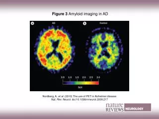
Conclusions • In vivo imaging is of use in the early identification of brain changes associated with AD. • The patterns of imaged brain changes appear to first involve the entorhinal cortex, then the hippocampus, and later the neocortex.