Download
1 / 30
320 likes | 401 Vues
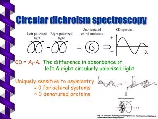
Circular Dichroism. Circular Dichroism?. 원편광 이색성 CD spectroscopy measures differences in the absorption of left-handed polarized light versus right-handed polarized light which arise due to structural asymmetry 분광학 (Spectroscopy)

E N D
Circular Dichroism? • 원편광 이색성 • CD spectroscopy measures differences in the absorption of left-handed polarized light versus right-handed polarized light which arise due to structural asymmetry • 분광학 (Spectroscopy) • 물질과 전자기파(빛)의 상호작용을 연구, 물질에서 방출되거나 물질에 흡수되는 스펙트럼을 분석하여 물질을 분석
Circular Dichroism? • 단백질 거울상(chiral) 이상질체인 D form과 L form으로 구성 • D/ L form이 동일한 양으로 구성된 화합물을 Racemic mixture • 편광된 빛이 asymmetric medium을 통과하면 optical rotation과 polarization loss 일어남 • Ellipticity(loss)를 측정하여 ellipticity 크기와 경향을 diagram으로 나타낸 것이 Circular Dichroism
Light • As an electromagnetic wave • Electrical component • Magnetic component • Direction • Two component는 서로 수직 • 빛의 진행방향에도 서로 수직
Polarization(편광) • 진행방향에 대해 수직으로 진동= 횡파의 특성 • 횡파의 경우, 진동방향이 파동의 진행방향의 수직인 평면상에 놓여있게 되어벡터의 성격을 갖게됨 • 편광 = 파동의 진동이 그 평면상의 특정 방향으로 놓이게 되는 것 • 편광방향 = 진동방향(물질에 더 큰 영향을 주는 전기장의 진동방향)
Polarization(편광) • 빛은 짧은 길이의 무수히 많은 파동줄기(wave train)가 모여서 형성된 것이므로 하나하나의 편광상태가 어떻게 집합되어 있는가를 고려해야 하므로 통계적인 처리가 필요 • 빛은 물질과 반응하여 편광상태가 바뀔 수 있음
Polarization(편광) • 선형편광 = 편광방향이 바뀌지 않음 • 전기장이 x 방향으로 진동하는 경우를 x 선형편광, y 방향 진동의 경우 y 선형편광 • x, y 선형편광을 적절히 조합하면 임의의 방향의 선형편광이나, 원형편광도 만들어 낼 수 있다.
Polarization(편광) • 전기장의 방향은 나선 형태로 계속 변함 • z 축 상의 한 점에 정지 한 채로 보면 다가오는 빛의 전기장은 점차 반 시계방향으로 회전을 하게 되는 것을 알 수 있다(좌향-원형편광)
Dichroism • The difference between the absorption of left and right handed circularly-polarized light is measured as a function of wavelength
Principle of CD • Chiral molecules은 좌원편광, 우원편광 된 빛을 다르게 흡수 ->CD spectrum • Chiral molecules은 optical active • Optical activity= 편광 된 빛이 물질을 통과할 때 편광 면을 회전시키는 성질
(a) Linear polarized light can be viewed as a superposition of opposite circular polarized light of equal amplitude and phase • (b) Different absorption of the left- and right hand polarized component leads to ellipticity (CD) and optical rotation (OR) (a) (b)
is therefore the angle between the initial plane of polarization and the major axis of the ellipse of the resultant transmitted light • A quantity is defined such thattan is the ratio of the major and minor axis of the ellipse of the transmitted light • ’ approximates the ellipticity • When expressed in degrees, ’ can be converted to a specific ellipticity [] or a molar ellipticity [] • CD is usually plotted as []
스펙트럼 • 자외선 가시광선에 의해 분자나 원자가 에너지를 흡수하여 전이를 일으켜 발생 • 흡수된 빛의 파장은 낮은 에너지준위의 전자를 높은 에너지 준위로 이동시킬 수 있는 에너지가 있음
전자전이 • 결합분자 궤도 함수(bonding moleculer orbital): , (에너지 준위가 낮음) • 반 결합 분자궤도 함수(antibonding moleculer orbital): *, * (에너지 준위가 높음) • 비결합 전자 (nonbonding electron): n 전자 (유기 화합물 중 O, N, S 및 할로겐 원자) • 전자의 전이는 전자가 결합성 궤도 함수(bonding orbital)에서 반결합성 궤도 함수(anti-bonding orbital)로 옮아가는 것을 의미함
Application • Near UV CD (250 - 350 nm) • Near-UV CD spectroscopy is dominated by Phe, Tyr, Trp and disulfides • When an aromatic residue is held rigidly in space, its environment is asymmetric, and it will exhibit CD
Application • Far UV CD (180 - 250 nm) • The amide group is the most abundant CD chromophore in proteins • In secondary structure conformations, the backbone and the amide bond chromophores are arranged in regular, organized, asymmetric patterns
Application • n -> π* centered around 220 nm • n -> π* involves non-bonding electrons of O of the carbonyl • π -> π* centered around 190 nm • π -> π* involves the π-electrons of the carbonyl • The intensity and energy of these transitions depends on φ and ψ (i.e., secondary structure)
Application • Far UV-CD of random coil: • positive at 212 nm (π->π*) • negative at 195 nm (n->π*) • Far UV-CD of β-sheet: • negative at 218 nm (π->π*) • positive at 196 nm (n->π*) • Far UV-CD of α-helix: • Exciton coupling of the π->π* transitions leads to positive (π->π*)(perpendicular) at 192 nm and negative (π->π*)(parallel) at 209 nm • negative at 222 nm is red shifted (n->π*)
Application • Determination of secondary structure of proteins that cannot be crystallized • Investigation of the effect of e.g. drug binding on protein secondary structure • Studies of the effects of environment on protein structure • Study of ligand-induced conformational changes
Application • 광우병 진단 • 정상적인 cellular prion protein 의 경우, 단백질의 2차 구조가 대부분 alpha-helix로 이루어져 있다. • 반면, 광우병에 걸린 scrapie prion protein은 beta structure 가 대부분의 2차 구조 구성 성분으로 변하게 된다. • Alpha helical structure 에서 beta-structure 로의 변화를 CD를 통해 확인할 수 있고, 광우병의 발병유무를 측정가능하다.
Sample Preparation • Additives, buffers and stabilizing compounds: Any compound which absorbs in the region of interest (250 - 190 nm) should be avoided. • A buffer or detergent or other chemical should not be used unless it can be shown that the compound in question will not mask the protein signal.
Sample Preparation • Protein solution: From the above follows that the protein solution should contain only those chemicals necessary to maintain protein stability, and at the lowest concentrations possible.Avoid any chemical that is unnecessary for protein stability/solubility. The protein itself should be as pure as possible, any additional protein or peptide will contribute to the CD signal.
Sample Preparation • Contaminants: Unfolded protein, peptides, particulate matter (scattering particles), anything that adds significant noise (or artifical signal contributions) to the CD spectrum must be avoided. Filtering of the solutions (0.02 um syringe filters) may improve signal to noise ratio. • Data collection: Initial experiments are useful to establish the best conditions for the "real" experiment. Cells of 0.5 mm path length offer a good starting point.
Sample Preparation • Nitrogen purging: the function of purging the CD instrument with nitrogen is to remove oxygen from the lamp housing, monochromater, and the sample chamber. The reason for removing oxygen is that oxygen absorbs deep UV light, thus reducing the light available for the measurement.
Sample Preparation • Typical Initial Concentrations: • Protein Concentration: 0.5 mg/ml • Cell Path Length: 0.5 mm • Stabilizers (Metal ions, etc.): minimum • Buffer Concentration : 5 mM or as low as possible while maintaining protein stability • Need very little sample: 0.1mg
Advantage • Simple and guick experiment • No extensive preparation • Measurements on solution phase • Relatively low concentrations/amounts ofsample • Microsecond time resolution • Any size of macromolecule
Disadvantage • Difficult to quantitate similarity or differences • Certain buffer components absorb strongly in Far-UV & can cause interference
Conclusion • 편광된 빛이 asymmetric medium을 통과하면 optical rotation과 polarization loss 일어남 • Ellipticity(loss)를 측정하여 ellipticity 크기와 경향을 diagram으로 나타낸 것이 Circular Dichroism • Near UV CD (250 - 350 nm) • Near-UV CD spectroscopy is dominated by Phe, Tyr, Trp and disulfides
Conclusion • Far UV CD (180 - 250 nm) • In secondary structure conformations, the backbone and the amide bond chromophores are arranged in regular, organized, asymmetric patterns • Random coil, β-sheet, α-helix