Download
1 / 61
610 likes | 619 Vues
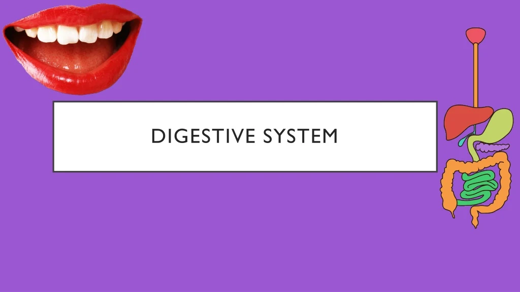
Digestive system. Organization of the digestive system. Digestive system has two categories of organs: those of the gastrointestinal tract and the accessory digestive organs Organs of the gastrointestinal tract Form a continuous tube Includes Oral cavity and pharynx Esophagus and stomach

E N D
Organization of the digestive system • Digestive system has two categories of organs: those of the gastrointestinal tract and the accessory digestive organs • Organs of the gastrointestinal tract • Form a continuous tube • Includes • Oral cavity and pharynx • Esophagus and stomach • Small intestine, large intestine, and anus • Within the lumen (inner opening), food broken down into smaller components to be absorbed
Accessory digestive organs • Assist in breakdown of food • Some producing secretions that empty into the GI tract • Include • Salivary glands, liver, pancreas • Teeth and tongue • Participate in chewing and swallowing • Gallbladder • Concentrates and stores liver secretions
General functions • Six main functions • Ingestion • Introduction of solid and liquid nutrients into the oral cavity • First step in process of digesting and absorbing nutrients • Motility • Voluntary and involuntary muscular contractions • Mixing and moving materials through the GI tract • Secretion • Process of producing and releasing fluid products facilitating digestion • E.g., digestive enzymes, acid, bile
Six main functions (continued) • Digestion • Breakdown of ingested food into smaller structures • Mechanical digestion • Material physically broken down by chewing and mixing • Chemical digestion • Involves specific enzymes to break chemical bonds • Change large complex molecules into smaller molecules • Absorption • Transport of digested molecules, electrolytes, vitamins, water • Move from GI tract into blood or lymph • Elimination • Expulsion of indigestible components that are not absorbed
GI TRACT WALL • GI tract • Hollow tube of 4 concentric layers, tunics • Innermost to outermost: • Mucosa • Submucosa • Muscularis • Adventitia (or serosa)
Mucosa • Inner-lining mucous membrane • Epithelium is in contact with lumen contents • Simple columnar epithelium allowing for secretion and absorption • Portions that must withstand abrasion (i.e., esophagus) are lined by nonkeratinized, stratified squamous epithelium • Underlying lamina propria • Composed of areolar tissue, small blood vessels, and nerves • Absorption occurs when substances move into these vessels • Smooth muscle deep to lamina propria: muscularis mucosae • Contractions facilitate release of secretions into lumen, increase contact of materials with mucosa
Submucosa • Areolar and dense irregular CT • Blood vessels, lymph vessels, nerves • Submucosal nerve plexus • Nerves and ganglia innervating smooth muscle and glands • Houses mucosa-associated lymphatic tissue (MALT) • Prevents ingested microbes from crossing GI tract wall • Peyer patches • Larger aggregates of lymphatic nodules in distal small intestine
Muscularis • Inner circular layer of muscle • Smooth muscle cells oriented circumferentially within GI tract • Contraction constricts tube lumen • Outer longitudinal layer • Cells oriented lengthwise along GI tract • Contraction shortens tube • Myenteric nerve plexus • Axons and ganglia between layers control contractions
Muscularis (continued) • Enteric nervous system • Submucosal plexus and myenteric plexus • Detects changes in tract wall and chemical makeup of lumen content • Sensory and motor neurons • Thickened at several points to form a sphincter • Closes off the lumen • Controls movement of materials into next section of GI tract
Muscularis (continued) • Functions to mix and propel contents within GI tract • Mixing • “Backward-and-forward” motion that lacks directional movement • Blends ingested materials with secretions • Propulsion • Directional movement of materials through GI tract • Occurs by peristalsis: sequential contraction of muscularis, GI tract wall moves like a wave
Adventitia or serosa • Outermost tunic may be either, depending on organ location • Adventitia • Areolar CT • Found outside the peritoneal cavity • Serosa • Same composition as adventitia • Completely covered by visceral peritoneum • Found within peritoneal cavity
Regulation of the digestive system • Enteric nervous system (ENS) • Sensory and motor neurons within submucosal plexus and myenteric plexus • Innervates smooth muscle and glands of GI tract • Coordinates mixing and propulsion reflexes • Autonomic Nervous System (ANS) • Parasympathetic innervation promotes GI tract activity • Sympathetic innervation opposes GI tract activity
Nerve reflexes • Baroreceptors detect stretch in GI tract wall • Chemoreceptors monitor chemical contents in lumen • Reflexes (by ANS or ENS) are initiated in response to receptor input • Short reflex – local reflex, only involves ENS; coordinate small segments of GI tract • Long reflex – involves sensory input to CNS and autonomic motor output; coordinate GI tract motility, secretions, and accessory digestive organs • Hormonal control • Several hormones participate in regulation of digestion • E.g., Gastrin, secretin, cholecystokinin, motilin
Gi tract organs • Upper GI tract organs and accessory structures • Oral cavity and salivary glands • Mechanical digestion begins • Saliva secreted from salivary glands in response to food • Contains salivary amylase, enzyme initiating digestion of starch • Mixed with ingested materials to form bolus • Pharynx • Bolus moved to pharynx during swallowing • Mucus secreted to facilitate swallowing
Upper GI tract organs and accessory structures (continued) • Esophagus • Bolus transported from pharynx into stomach • Lubricated by mucus secretions • Stomach • Bolus mixed with gastric secretions by smooth muscle contractions • Secretions produced by epithelial cells of stomach • Chyme formed from mixing • Duodenum also considered part of upper GI tract
Oral cavity • Mouth, entrance to GI tract • Two distinct regions: vestibule and oral cavity proper • Vestibule • Space between gum, lips, cheeks • Oral cavity proper • Lies central to the teeth • Leads posteriorly into oropharynx • Cheeks, contains buccinator muscles • Compress cheeks to hold solid material while chewing
ORAL CAVITY & SALIVARY GLANDS • Salivary glands • Produce saliva • Intrinsic salivary glands (within oral cavity) • Unicellular glands • Continuously release secretions independent of food • Contains lingual lipase, enzyme that begins digestion • Extrinsic salivary glands (outside of oral cavity) • Produce most saliva • Parotid, submandibular, and sublingual glands
Salivary glands (continued) • Parotid salivary glands, largest salivary glands • Anterior and inferior to ear • 25–30% of saliva • Saliva conducted thorough parotid duct to oral cavity • Extends from gland across masseter, opening near second upper molar • Infection of the parotid glands causes mumps
Salivary glands (continued) • Submandibular salivary glands • Inferior to oral cavity floor and medial to mandible body • Produces 60–70% of salliva • Submandibular duct opens from each gland to floor of cavity • Sublingual salivary gland • Inferior to tongue and medial and anterior to submandibular glands • Extends tiny ducts opening into inferior surface of cavity • Contribute only 3–5% of saliva
HISTOLOGY: SALIVARY GLANDS • Two types of secretory cells within salivary glands • They produce the components of saliva: mucous cells and serous cells • Mucous cells • Secrete mucin, forming mucus upon hydration • Serous cells • Secrete watery fluid containing electrolytes and salivary amylase • Proportion of each varying among glands
Saliva • 1.0–1.5 L secreted daily, most produced during mealtime • 99.5% water and a mixture of solutes • Salivary amylase, mucin, lysozyme added • Functions of saliva: • Moistens ingested food to help become bolus • Salivary amylase initiates chemical breakdown of starch • Food molecules dissolved here so taste receptors stimulated • Cleanses oral cavity structures • Antibacterial substances inhibit bacterial growth (lysozyme, IgA antibodies)
Mechanical digestion: mastication • Mastication, chewing • Mechanically reduces bulk to facilitate swallowing • Increases surface area to facilitate exposure to digestive enzymes • Promotes salivation • Requires coordinated activities of teeth, lips, tongue, cheeks, jaws • Controlled by nuclei in medulla and pons, mastication center
Teeth • Collectively known as the dentition • Exposed crown andconstricted neck • One or more roots, anchoring it to jaw • Fit tightly into dental alveoli, sockets within alveolar processes • Bound to processes by periodontal ligament • Gomphosis joint: roots, dental alveoli, periodontal ligament
Deciduous and permanent teeth • 20 Deciduous teeth • Erupt between 6 and 30 months • 32 Permanent teeth, replacing deciduous teeth • More anteriorly placed permanent teeth appearing first • Third molars, wisdom teeth, in late teens or 20s • May emerge partially or become impacted
Deciduous and permanent teeth (continued) • Incisors, most anteriorly placed teeth • Shaped like chisel for slicing food • Canines, posterolateral to incisors • Pointed tip for puncturing and tearing food • Premolars, posterolateral to canines • Flat crowns with prominent ridges (cusps) that crush and grind • Molars, most posteriorly placed teeth • Large, broad crowns and cusps • Grinding and crushing materials • In each quadrant • 2 incisors, 1 canine, 2 premolars, 3 molars • Gingivae,gums • Dense irregular CT • Overlying nonkeratinized stratified squamous epithelium • Covers alveolar processes and surrounds neck of teeth
Pharynx & esophagus • Gross anatomy of the pharynx • Funnel-shaped muscular passageway • Passageway for air and food • Formed by 3 skeletal muscle pairs • Superior, middle, and inferior pharyngeal constrictors • Lined with nonkeratinized stratified squamous epithelium • Protection against abrasion
Gross anatomy of the esophagus • Esophagus: normally collapsed, tubular passageway • Begins at level of cricoid cartilage • Directly anterior to vertebral bodies • Inferior region connecting to the stomach • Passes through opening in diaphragm, esophageal hiatus • Last 1.5 cm in abdomen
Motility: the swallowing process • Swallowing,deglutition • Moving ingested materials from oral cavity to stomach; 3 phases • Voluntary phase, occurring after ingestion • Controlled by cerebral cortex • Bolus formed as ingested materials and saliva mix • Bolus directed posteriorly toward oropharynx • Pharyngeal phase • Involuntary reflex • Tactile sensory receptors around fauces stimulated • Initiate sensory input to swallowing center in medulla oblongata • Signals relayed to effectors
Motility: the swallowing process (continued) • Effector response of pharyngeal phase • Entry of bolus into oropharynx • Elevation of soft palate and uvula to block passageway between oropharynx and nasopharynx • Elevation of larynx by extrinsic muscles • Move epiglottis to cover laryngeal opening • Prevents ingested material from getting into trachea • Nerve signals sent to medulla oblongata to ensure breath not taken during swallowing
Motility: the swallowing process (continued) • Esophageal phase • Involuntary phase when bolus passes through esophagus • Bolus stimulates sequential waves of muscular contraction • Propels bolus toward stomach • Superior and inferior esophageal sphincters closed at rest • Relax when bolus swallowed • Contract again afterwards, preventing reflux of materials
STOMACH • Located in superior left abdominal quadrant, inferior to diaphragm • Chemical and mechanical digestion continues in stomach • Digestion of protein and fat begins in stomach • Ingested materials spending 2 to 6 hours here • Absorption limited to small, nonpolar substances • Serves as “holding bag” for controlled release of partially digest material
Gastric secretions • Produced by 5 types of secretory cells • 4 produce gastric juice, fifth type secretes hormone • Surface mucous cells • Line stomach lumen and extend into gastric pits • Continuously secrete alkaline product containing mucin • Mucous layer helps to prevent ulceration of stomach lining • Protects from gastric enzymes and high acidity
Gastric secretions(continued) • Mucous neck cells • Produce acidic mucin • Help maintain acidic conditions • Both types of mucous cells help protect the stomach lining from abrasion and injury
Gastric secretions (continued) • Parietal cells—add two substances to stomach • Intrinsic factor • Required for absorption of vitamin B12 in ileum • Necessary for production of normal erythrocytes • Hydrochloric acid • Forms from H+ and Cl– secreted across cells’ surface • Responsible for low pH of stomach • Hydrochloric acid functions • Helps break down plant cells walls and animal CT • Denatures proteins, facilitating chemical digestion • Converts inactive enzyme pepsinogen into active pepsin • Kills most microorganisms entering stomach
Gastric secretions (continued) • Chief cells • Most numerous secretory cells within gastric glands • Produce and secrete packets of zymogen granules • Primarily containing pepsinogen, inactive precursor of pepsin • Pepsin must be in inactive form to prevent destruction of chief cell proteins • Pepsinogen activated by HCl and other active pepsin molecules • Chemically digests denatured proteins into oligopeptides • Produce gastric lipase, playing limited role in fat digestion (digests about 10-15% of ingested fat)
Stomach motility performs two primary functions: mixing the bolus to form chyme and emptying chyme from stomach to small intestine • Gastric mixing • Form of mechanical digestion • Changes semidigested bolus into chyme • Churned and mixed, leading to reduction in size of swallowed particles
Motility in the stomach (continued) • Gastric emptying • Movement of acidic chyme from stomach into duodenum • Pressure gradient moving contents toward pylorus • Gradient increasing force against pyloric sphincter • Sphincter opens, with entrance of small volume of chyme • Sphincter closes, with retropulsion • Reverse flow of some contents back toward stomach
Overview of the gi organs • Lower GI tract organs • Process of digestion and absorption continues • Elimination of indigestible and unabsorbable material • Small intestine • Divided into 3 continuous regions: duodenum, jejunum, ileum • Duodenum, part of upper GI tract • Receives chyme from stomach mixed with accessory organ secretions • Most chemical digestion and absorption happens here
Lower GI tract organs (continued) • Accessory digestive organs • Secretions of bile and pancreatic juice • Bile produced by liver • Stored, concentrated, released by gallbladder • Pancreatic juice with digestive enzymes secreted from pancreas • Large intestine • Primarily absorbs water, electrolytes, some vitamins • Feces produced and eliminated through anus