Download
1 / 64
660 likes | 690 Vues
ANAEROBIC BACTERIA Anaerobic Gram positive rods. Doç.Dr.Hrisi BAHAR Istanbul University Cerrahpasa Medical Faculty. ANAEROBIC GRAM POSITIVE RODS. 1 * SPOROGENOUS ANAEROBIC GRAM POSITIVE RODS 2 * ASPOROGENOUS ANAEROBIC GRAM POSITIVE RODS. * SPOROGENOUS ANAEROBIC GRAM POSITIVE RODS.

E N D
ANAEROBIC BACTERIAAnaerobic Gram positive rods Doç.Dr.Hrisi BAHAR Istanbul University Cerrahpasa Medical Faculty
ANAEROBIC GRAM POSITIVE RODS 1*SPOROGENOUS ANAEROBIC GRAM POSITIVE RODS 2*ASPOROGENOUS ANAEROBIC GRAM POSITIVE RODS
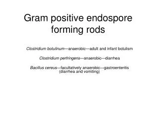
*SPOROGENOUS ANAEROBIC GRAM POSITIVE RODS CLOSTRIDIA ●Generally large rods that are often club shaped due to presence of spore. ● Sporulation is variable ●All initially stain gram positive but may become gram negative rapidly.
Pathogenic Potential of Clostridium-1- ●Very proteolytic and aerogenic; elaborate multiple toxins. ● Common inhabitants of the gastrointestinal tract and the female genital tract.
Pathogenic Potential of Clostridium-2- Some members associated withspecificdiseases. ● I.Gas Gangrene C. perfringens (type A), C. septicum C novyi type A, C. bifermentans C. histolyticum, C. sordelli, C. sporogenes ● II.Tetanus‑ C. tetani ● III.Botulism ‑ C. botulinum
Pathogenic Potential of Clostridium-3- ● IV.Antibiotic-associated Diarrhea and Colitis C. difficile; rare C. perfringens type C ● V.Miscellaneous infections - wounds, abscess, soft tissue infection, myonecrosis, bacteremia: C. perfringens, C. ramosum, C. septicum, C. sporogenes and others
Tetanus-1- • The spores of C. tetani are found in feces of humans and animals and in soil. *Signs and symptoms :Tetanus affects skeletal muscles. * Mortality rates vary from 40% to 78%.
Tetanus-2- ●Generalized tetanusis the most common type of tetanus, representing about 80% of cases. ●Neonatal tetanusis a form of generalized tetanus that occurs in newborns.
Tetanus-3- Neonatal tetanus ● A frequent cause of death in developing countries ● Most common causes: cutting the umbilical cord with unsterilized instruments or infection of the umbilical stump ●The fatality rate: around 90% ● The common death cause: respiratory failure
Tetanus-4- ● Local tetanus Uncommon form of the disease ●Cephalic tetanusis a rare form of the disease
Tetanus-5- The tetanus toxin (tetanospasmin) travels to the central nervous system and binds to nerve tissue. Prevents the inhibitory signals to the motor neurons and results in prolonged contraction of extensor and flexor muscles. C. tetani also produces tetanolysin.
Tetanus-6- ●Muscle spasms mediated by tetanospasminproduces a characteristic arching posture known as opisthotonos. ●Without treatment, the patient ultimately succumbs to respiratory failure. ●The incubation period ranges from 1‑54 days; generally 6‑15 days.
Tetanus-7- Opisthotonos
Tetanus-8- Diagnosis ● The diagnosis is based on clinical symptoms and patient history (i.e.lack of current vaccination). ●No attempt is made to cultivate the organism. ●A Gram stain of wound exudate (if available) may demonstrate Gram positive rods and gram negative rods with swollen ends due to the presence of spores.
Tetanus-9- Latent period: 4-5d ~ several weeks Lockjaw, sardonic smile opisthotonos
Tetanus -11- Therapy Treatment usually focuses on ● controlling muscle spasms, ● stopping toxin production and ● neutralizing the effects of the toxin.
Botulism -1- ●The causative agent, C. botulinum, is found in soil, freshwater, and seawater sediments. ● Botulism is a neuroparalytic disease. ● The toxin of C.botulinum acts by blocking nerve function and leads to respiratory and musculoskeletal paralysis. ● The toxin affects nerves,it affect specifically, the inhibition of the release of acetylcholine from motor neurons.
Botulism -2- ● Botulism toxin is one of the most powerful known toxins: about one microgram is lethal to humans. ● In all cases illness is caused by the toxin made by C. Botulinum.
Botulism-4- Mode of acquisition 1-Infant botulism. ●The most common form in Western countries , ●This occurs in small children who are colonised with the bacterium during the early stages of their life. ●The bacterium releases the toxin into the intestine, the consumption of honey during the first year of life has been identified as a risk factor for infant botulism.
Botulism-5- 2-Foodborn botulism. Results from contaminated foodstuffs in which C. botulinum spores have been allowed to germinate and produce botulism toxin. 3-Wound botulism. Results from the contamination of a wound with the bacteria
Botulism-6- Diagnosis ●C.botulinum infections should be made on clinical grounds. ● Confirmation of the diagnosis is made by testing of a stool or enema specimen with the mouse .
Botulism-8- Treatment * The only drug currently available to treat infant botulism is “Botulism Immunoglobulin Intravenous Human”. * If diagnosed early, foodborne and wound botulism can be treated by inducing passive immunity with a “horse-derived antitoxin”.
Pseudomembranous colitis-1- ● Infection of the colon,often,but not always caused by the bacterium C.difficile. ●This disease is induced by long term use of broad spectrum antibiotics ● It is characterized by the presence of colonic plaques that form a pseudomembrane.
Pseudomembranous colitis-2- ● Key factor in the pathogenesis of PMC is a disturbance of the normal bowel flora that permitsC. difficile to overgrow the endogenous flora, produce toxin and induce disease. ●Approximately 2% of healthy adults carry C. difficile as commensal flora
Pseudomembranous colitis-3- Diagnosis ●Visualization of characteristic pseudomembranous plaques by colonoscopy ● Detection of the toxin is the most accurate diagnosis way but is difficult to perform. ●C. difficile can be cultured on special media CCFA‑cycloserine, cefoxitin, egg yolk, fructose agar.
Pseudomembranous colitis-4- MAP :Mitojen Activated Protein Kinases
Pseudomembranous colitis-6- ● The disease is usually treated by oral metronidasole (500 mg every 8 hours) ● Oral vancomycin (125 mg every 6 hours) ● Additionally treatment by probiotics
Anaerobic cellulitis-1- ● Invasion of necrotic wound tissue by the proteolytic clostridia. ●Characterized by gas accumulation, discoloration of the underlying skin, and the presence of a malodorous, brownish, purulent discharge.
Clostridial myonecrosis or gas gangrene-1- ● Gas gangrene is associated with severe deep wounds. ●Clostridial myonecrosis or Gasgangrenedevelop in a lowered oxidation‑reduction potential present in the wound.
Clostridial myonecrosis or gas gangrene-2- This situation was caused by ●The presence of foreign bodies in the wound, ● Failure of the blood supply to the infected area, ● The presence of necrotic tissue and hemorrhagie in the wound, ● The presence and multiplication of other bacteria.
Clostridial myonecrosis or gas gangrene-3- ● Clinical features of gas gangrene include marked systemic toxicity, fever,tachycardia, and a tender, painful edematous wound with a sweet‑ or foul‑smelling discharge . ● The most frequently encountered species are C. perfringens (predominant), C. novyi and C. septicum, and Clostridium clostridiforme.
C. perfringens food-borne illness -1- ● This is reported to be the third most common form of food poisoning in the world. ● The predisposing factor appears to be the ingestion of improperly cooked/stored meat or meat product. ● Most patients are afebrile.
C. perfringens food-borne illness -2- ● There is nausea and vomiting in one third of the cases; stools are usually foamy and foul smelling. ● The illness is usually mild and self-limiting. ●Diagnosis is confirmed by the recovery of a large number of organisms (>108 spores/g feces) from stool.
Isolation and Identification of Clostridia -1- ●Identification of the commonly encountered clostridia can often be accomplished on the basis of colony morphology, growth on selective media, aerotolerance determination and a few screening tests. ●Egg Yolk agar (EYA) is commonly employed for the identification of clostridia.
Isolation and Identification of Clostridia -2- ●EYA is useful for the detection of two enzymes: lecithinase ‑ precipitation around colony on EYA,lipase‑ iridescent sheen at edge of colony on EYA. ● Alpha toxin is a specific form of lecithinase produced by C. perfringens and a few other clinically important clostridia.
NAGLER POSITIVE CLOSTRIDIA • Clostridium perfringens • Clostridium sordelii • Clostridium bifermentans
ASPOROGENOUSANAEROBIC GRAMPOSITIVE RODS ●Heterogeneous group of organisms that may be recoveredas commensal floraon a variety of mucosal surfaces including the: upper respiratory tract, gastrointestinal tract, female genitourinary tract.
ASPOROGENOUS ANAEROBIC GRAM POSITIVE RODS -1- ● The genera observed vary with the anatomic site. These organisms are considered to be opportunistic pathogens that take advantage of breaks in the integrity of the mucosal surfaces. ●Usually recoveredfrom polymicrobic infections involvingfacultative bacteria (enterics,staphylococci,streptococci) or anaerobic gram negativerods (Bacteroides or Fusobacterium).
ASPOROGENOUS ANAEROBIC GRAM POSITIVE RODS -2- The most commonly encountered genera are: ● Actinomyces, ● Bifidobacterium, ● Eubacterium, ● Propionibacterium, ● Mobiluncus, ● Lactobacillus
Actinomyces are branching anaerobic or microaerophilic , non-sporulating Gram positive bacilli ACTINOMYCES
ACTINOMYCOSIS -1- ● Actinomycosis is an endogenous infection caused byActinomyces sp. ● The infection usually manifests as a chronic suppurative disease that may produce granulomatous lesions. ● The infection spreads by direct extension through contiguous tissue including connective tissue and bone .
ACTINOMYCOSIS -4- Clinical manifestations include ● Cervicofacial ● Thoracic ● Abdominal ● Skin lesions ● Periodental diseases ● Pelvic infections.
BIFIDOBACTERIUM Pathogenicity Members of this genus are oral and intestinal commensal flora which are associated with polymicrobic infections, especially pulmonary. They are rarely encountered in clinical material. B. dentium appears to be the only species with pathogenic potential.