Download
1 / 14
140 likes | 161 Vues
Myopathies. Madison Pilato. Myopathy. Case Congenital Myopathies Critical Illness Myopathy Inflammatory Myopathies References. Case. 68 yo M w/o significant medical history p/w slowly progressive muscle weakness for 5 years. First symptom R knee giving out on stairs

E N D
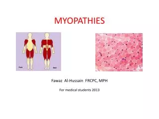
Myopathies Madison Pilato
Myopathy • Case • Congenital Myopathies • Critical Illness Myopathy • Inflammatory Myopathies • References
Case 68 yo M w/o significant medical history p/w slowly progressive muscle weakness for 5 years. First symptom R knee giving out on stairs Today has difficulty rising from a chair and grasping with right hand Exam: Intact CN, sensation and DTRs RUE: 5/5 shoulder abduction, 5/5 elbow flexion and extension, 4/5 wrist flexion 5/5 wrist extension, 3-/5 finger flexion LUE: 4+/5 finger flexion, rest is 5/5 RLE: 4+/5 knee extension, rest 5/5 LLE: 4+/5 hip flexion, 3+/5 knee extension, 4+/5 dorsiflexion Case from Continuum, “Pattern Recognition Approach to Myopathy” Volume 12, Issue 3, p13-32
https://adc.bmj.com/content/88/12/1051 Core MyopathyCentral core disease • Clinical spectrum: apparent normalcy to severe weakness • Typically: hypotonia in infancy, developmental delay in childhood • Proximal limbs, axial muscles w/musculoskeletal abnormalities (hip dislocation, kyphoscoliosis, joint contractures) • Autosomal dominant • RYR1 mutation – majority of cases, rare resp weakness, rare cardiomyopathy. Malignant hyperthermia • SEPN1 severe respiratory weakness, severe scoliosis • Pathology: • Fibers w/regions devoid of oxidative enzyme activity • Predominance of type 1 muscle fibers RYR1 Malignant hyperthermia
https://www.sciencedirect.com/science/article/pii/S0960896613009942#f0005https://www.sciencedirect.com/science/article/pii/S0960896613009942#f0005 Nemaline (rod) myopathy “Nema”=thread • Variable presentation; genetically heterogeneous (AD, AR) • Infantile hypotonia: prominent facial and respiratory weakness (classic presentation), severe form with 20% mortality • Adult-onset: head-drop, dysphagia, respiratory insufficiency • Pattern of weakness: facial muscles, neck/trunk flexors, dorsiflexors, toe extensors • Earlier onset usually more severe. Less severe forms can stabilize or even improve • Pathology: • Rod shaped structures within muscle fibers • atrophic fibers without grouping • Trichrome stain: reddish purple thread-like inclusions within sarcoplasm • Type 1 fiber predominance • Ultrastuctural – intracytoplasmic rod-shaped structures
Centronuclear MyopathyMyotubular Myopathy • Clinically and genetically heterogeneous • Muscle fibers with large central nuclei that resemble myotubes (early fetal muscle fibers) • Severe, more common form is X linked. Occurs in male infants, marked hypotonia and weakness, respiratory muscle weakness and failure. Facial weakness and impaired bulbar function. Female carriers can have limb girdle or facial weakness. 1/3 die from respiratory complications in infancy • Less common form is AD or AR. Mild weakness and hypotonia may be unrecognized. • Pathology: • One or more centrally placed nuclei with surrounding clear area • Persistent fetal characteristics of muscle
Steroids Neuromuscular Blockade Severe systemic illness Critical Illness MyopathyAKA acute quadriplegic myopathy • Critically ill patient cannot be weaned off of ventilator, diffuse flaccid paralysis • 2/3 conditions usually required: corticosteroid therapy, neuromuscularblockade, severe systemic illness (e.g., sepsis) • ~25% of critically ill patients on MV longer than 1 week • Rapid onset, proximal weakness; may be asymmetric, may be muscle wasting • More common females • Resolves 3 weeks to one year; stop any offending agents • Pathology: • atrophy of muscle fibers (non-specific) • Electronic microscopic features are diagnostic/pathognomonic, selective loss of thick (myosin) filaments
http://n.neurology.org/content/66/1_suppl_1/S20.figures-only Inclusion body myositis • Most common primary muscle disease over age 50; 3:1 male predominance • Slow symptom progression; asymmetric • Early involvement of quadriceps and ankle dorsiflexors; frequent falls • Early involvement wrist and finger flexor muscles • ~50% swallowing difficulties; ~1/3 facial weakness • Muscle atrophy can be prominent • Can include peripheral polyneuropathy and sensory complaints • Poorly responsive to immunosuppressive tx, 50% WC bound after 10 years • Tx: supportive, exercise, supplement with creatine monohydrate • Pathology: • Intracytoplasmic vacuoles rimmed with granular material; inclusions react w/Abs associated with neurodegenerative diseases including beta-amyloid, tau, ubiquitin • Ultrastructual: diagnostic feature, inclusions w/in muscle cytoplasm, nuclei or both
Dermatomyositis= small vessel vasculitis • Proximal muscle weakness, weeks to months • Characteristic rash: red rash over eyelids and extremities (heliotrope, shawl, Gottrons) • Humorally mediated microangiopathy/vasculitis with ischemic necrosis of muscle fibers • Most common inflammatory myopathy of childhood, seen in all ages • 3:2 female predominance • Frequency of cancer increased for 3 years after onset in adults • Tx: immunosuppressive • Pathology: • Inflammation of perimysialblood vessels • Lymphocytes plus plasma cells and eosinophils (mixed infiltrate) • Necrosis and phagocytosis in groups (microinfarcts) • Perifascicular atrophy = sublethal myofiber stress @ distal end of blood supply
Polymyositis • Controversial; diagnosis of exclusion • Proximal muscle weakness over weeks to months with elevated CPK without skin changes of dermatomyositis. 2:1 female predominance • Oculobulbar rarely affected • May be associated with other autoimmune/collagen vascular diseases • Tx with immunosuppressive therapies (corticosteroids); 1/3 pts left with disability • Pathology: • Lymphocytic inflammation surrounding and invading otherwise healthy-appearing muscle fibers. • Rimmed vacuoles (IBM) and other types of inflammatory cells should be absent • CD-8 positive T cells, muscle fibers demonstrate upregulation MHC-I
References • Continuum, “Pattern Recognition Approach to Myopathy” Volume 12, Issue 3, p13-32 • Prayson Neuropathology, 2nd Edition, Skeletal Muscle and Peripheral Nerve Disorders • Up to date: Congenital Myopathies • Up to date: Clinical manifestations of dermatomyositis and polymyositis in adults