Download
1 / 20
200 likes | 325 Vues
1 Peterová K, 2 Brabec J, 3 Svobodová R, 3 Olejárová M, 3 Závada J, 3 Fojtíková M, 4 Potyšová Z, 4 Pešičková SS, 1 Vojtěchová A, 1 Peterová V, 2 Petrovický P. Amygdalar MR changes in systemic lupus patients.

E N D
1Peterová K, 2Brabec J, 3Svobodová R, 3Olejárová M, 3Závada J, 3Fojtíková M, 4Potyšová Z, 4Pešičková SS,1Vojtěchová A, 1Peterová V, 2Petrovický P. Amygdalar MR changes in systemic lupus patients 1MR Department, RadiodiagnosticClinic;2Institute of Anatomy; 3Institute ofRheumatology;4Department ofNephrology, FirstFacultyofMedicine, Charles University in Prague, U nemocnice 2, Prague 2, CzechRepublic.
Aim of the study • to analyze changes of amygdalae and their volumetry in NP-SLE* patients • patients with depression and mood disorders had been reported to have disability of amygdalae, changes in size and activity, which in some studies correlated with the severity of depressive episodes • NP-SLEsymptomsinclude cognitive deficits up to dementia, mood disorders, depression, anxiety, feelings of fear, strokes, epilepsy etc. *NP-SLE = systemic lupus erythematosus with neuropsychiatric symptomatology
Examination of patients • Clinical examination – neurological, rheumatolological • Neuropsychological investigation • Mini-Mental State Examination (MMSE), EEG • Neuropsychiatric Inventory (NPI) • Cerebral magnetic resonance (MR) • Complete laboratory tests including autoantibodies • The cumulative dose of corticoids were counted, the actual treatment was followed • The duration of SLE disease and the duration of SLE and NP symptomatology in years at the time of MRI • Pall patients were prospectively compared with corresponding healthy persons
Group of patients • 23 femalewith proved NP-SLE • Age: 19 to 67 years • Durationof SLE disease: 0.25 to 15 years • Durationof NP symptomatology: 0.25 to 12years • Activityofdisease (measured by SLEDAI) 0 – 26 • Patientsweremoderatelyfunctionallyimpaired • Allpatientsweretakingmedicationatthetimeofthe MR scan • psychotropicmedications (meannumber : 2.4 ± 1.0) • corticoids (cumulativedose: 480 - 89 400) • Serumlevelsof C-reactive protein: 0.3 – 45
Exclusion criteria for all patients • Other unstable illness • Medical illness, which could cause the neuropsychiatric symptoms (eg. multiple sclerosis, other rheumatological disease) • Pregnancy • Substance abuse within past 6 months
Controls • 23 healthy female controls • Mean age: 40.43 ± 22.3 years • Individually matched with patients for age • Investigated by clinical doctor and by MR only
Method • 1.5T scannerPhilips, head coil • T1 weighted images (T1WI) in FFE echo mode in transversal plane with TR 25ms, TE 5ms, FA 30o in 1mm thin slices without gap; • T2 weighted images (T2WI) in Turbo Spin mode in transversal and sagittal planes:TR 4540ms, TE 100ms, FA 90o,3mm thin slices; • Flow atenuation recovery mode(FLAIR)in transversal plan: TR 11000ms,TE 140ms,TI 2600, FA 90o,1.5mm thin slices without gap; • IV application of gadolinium to all investigated patients, • healthy controls were investigated only without contrast,
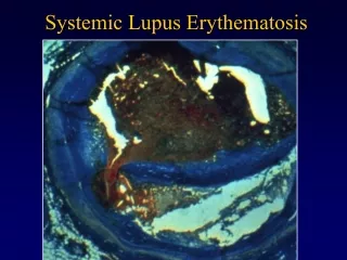
Histology – Nissl I. Classical amygdala Expanded amygdala
Volumetric procedure • included the following processing of each subject's brain image: • semi-automatic brain extraction (removal of skull and dura signal) • semi-automatic segmentation (i.e. separation of tissue into white or gray matter and cerebrospinal fluid); • automatic spatial transformation (transformation of tissue maps to stereotaxic space - Talairach model); • modulation (volume preservation that corrects the unintended changes in total tissue volume produced by spatial transformation).
Statistical analysis • Spearman´s non - parametric correlation coefficient • Wilcoxon´s non-parametric test for 2 dependent selections • Programs from the statistical software SPSS 13.0.
Results of our study • In group of patients with active NP-SLE • right amygdala correlated from all monitored parameters with only the left amygdala (p = 0.01) • left amygdala correlated with right amygdala (p = 0.01) and with volumetry in flow atenuation inversion recovery (FLAIR) and T1 weighted images (p = 0.05) • Volume of amygdalae did not correlate with total cerebral volume (in FFE, FLAIR), brain-parenchyme fraction (BPF), disease duration, time from first SLE and NP sympt., lesion load, laboratory or cognitive tests
Conclusions • We failed to demonstrate significant volume change in amygdalae • We did not demonstrate significant difference of amygdalar volume due to the duration of NP-SLE or age of the patients • The authors’ research was supported by the research project MZO 00064165,CEZ JI 3/98:11110001
ThankYouforyourattention We thank to all colleagues participating in the project: Prof. Zdeněk Seidl, M.D.PhD. Prof. Ctibor Dostál, M.D.DrSC. Prof.Vladimír Tesař, M.D. DrSC. Satu Sinikka Pešičková, M.D.PhD. Zuzana Potyšová, M.D. PhD Zuzana Abrahamová-Hladinová, M.D. PhD. Marta Olejárová, M.D. PhD. Jakub Závada, M.D. PhD Martina Fojtíková, M.D. PhD. Svobodová Radka, M.D. Tegzová Dana, M.D. PhD. Milada Lősterová Naděžda Baxová Antonie Vojtěchová Martina Strouhalová Věra Ečerová Miroslav Kron Jiří Brabec, M.D.