Download
1 / 58
620 likes | 871 Vues
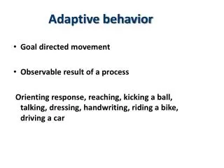
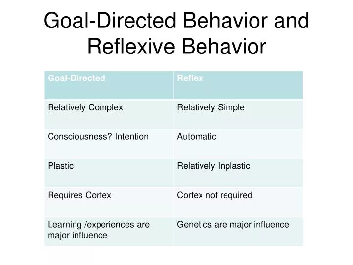
Goal-Directed Behavior and Reflexive Behavior. Goal-Directed Behaviors Require:. Goal selection and prioritization Resistance to distracters -Cross-modal Sensory integration Perception of target Awareness of location of movable body part Ability to aim movement of body part

E N D
Goal-Directed Behaviors Require: • Goal selection and prioritization • Resistance to distracters -Cross-modal Sensory integration • Perception of target • Awareness of location of movable body part • Ability to aim movement of body part • Ability to detect errors and re-adjust, (use feedback) • Ability to use feedback to control movement of body part
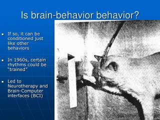
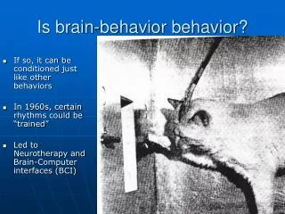
THE DLPFC: “The conductor” Integrates cross modal input- may initiate goal-directed behaviors Lesions of the dorsolateral frontal areas results in a number of “executive” motor impairments. These include perseveration, incoordination, motor impersistence, apraxias and hypokinesia. http://www.youtube.com/watch?v=p_uhP1vDfoo
The premotor and supplementary motor ctx: “The sections” Stimulation= complex sequences of behavior (aimless behavior)
Damage to the secondary Motor Cortex? • Ideomotor Apraxia • This apraxia is associated with great difficulty in the sequencing and execution of movements. A common test of apraxia is to request the patient to demonstrate the use of a tool or household implement (e.g., "Show me how to cut with scissors"). Difficulties are apparent when the patient moves the hand randomly in space or uses the hand as the object itself, such as using the forefinger and middle finger as blades of the scissors. They have additional trouble sequencing the correct series of movements and make errors in orienting their limbs in space consistent with the desired action. Imitation of the movements of others will usually improve performance but it is still usually defective. • Memories for skilled acts are probably stored in the angular gyrus of the parietal lobe in the left hemisphere. • http://www.youtube.com/watch?v=gewP1T7GYcc
The primary motor cortex; “the instrument” Stimulation = relatively simple fragments of behavior
TWO MAJOR DESCENDING PATHWAYS FROM THE PRIMARY MOTOR CORTEX: The Dorsolateral pathway
And the VM Path. • The VM pathway does not discretely decussate, but does branch and innervate contra lateral segments in the spinal cord.
Dorsolateral Decussates at medullary pyramids Distal muscle groups More direct More volitional control Higher resolution of control Ventromedial Does not cross Medial muscle groups Gives off spinal collaterals Yoking Lower resolution of control DL vs VM descending motor paths
Other Motor Pathways • In addition there are other motor paths that have relays in the brainstem • These other paths innervate nuclei of the RAS, cranial nerve nuclei, etc…
Amyotropic lateral sclerosis (ALS)disease of the alpha motor neurons
Alpha motor neurons project to form part of spinal nerve pairs
Muscle groups are complex; attach bone to bone via tendons and ligaments
The Neuromuscular junction (NMJ): The receptive portion of muscle-the motor end-plate
End-plate potential • Larger • Longer • Leads to Ca+ influx in sarcolema of muscle • Ca+ causes muscle contraction
muscle fibers encase myofibrils. The casing is called the sarcolema Muscle group myofibril Muscle fiber
When the NMJ is activated Actin-myosin interact to shorten the length of a muscle fiber
Goal-directed Complex Higher levels of control Plastic Numerous reflexive behaviors are involved Reflexive Simple Automatic inplastic Cortical vs Spinal control of behavior
Spinal reflex ARCs • Monosynaptic • stretch • Polysynaptic • Withdrawal • Antagonist muscle groups • Synergistic muscle groups • Polysegmental relexes • Cross-spinal reflexes
Stretch reflex regulates muscle tension in every muscle group
The polysynaptic part of stretch reflexes: inhibition of Antagonist muscles
Spinal inhibition of antagonist muscles require inhibitory interneurons