Download
1 / 24
240 likes | 375 Vues
The Axial Skeleton – part 2 The Vertebral Column. The spine or vertebral column : protects the spinal cord supports the head and body. 26 bones: 24 vertebrae , the sacrum, and coccyx. Regions Cervical (C) – 7 v Thoracic (T) – 12 v Lumbar (L) – 5 v Sacral (S) Coccygeal (Co).

E N D
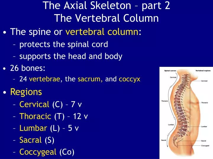
The Axial Skeleton – part 2The Vertebral Column • The spine or vertebral column: • protects the spinal cord • supports the head and body • 26 bones: • 24 vertebrae, the sacrum, and coccyx • Regions • Cervical (C) – 7 v • Thoracic (T) – 12 v • Lumbar (L) – 5 v • Sacral (S) • Coccygeal (Co)
Curvatures • Cervical curve • Thoracic curve • Lumbar curve • Sacral curve • Primary Curves • Thoracic and sacral curves - present during fetal development • aka accommodation curves-accommodate internal organs • Secondary Curves • Lumbar and cervical curves-appear after birth • Aka compensation curves-shift body weight for upright posture
Vertebrae • 3 Parts of a Vertebra • vertebral body (centrum)-transfers weight along the spine • vertebral arch-posterior margin of vertebral foramen • articular processes-lateral projections between laminae and pedicles
Intervertebral Discs • Are pads of fibrocartilage • Separate the vertebral bodies • Absorb shocks Figure 7–17d,e
Vertebral Regions • Vertebrae are numbered: • by region, from top to bottom • C1 articulates with skull, L5 with sacrum • Vertebrae of each region: • have characteristics determined by functions Figure 7–16
The Cervical Vertebrae • Characteristics of C1–C7: • small body (support only head) • large vertebral foramen (largest part of spinal cord) • C1 (atlas) has no spinous process all others have short spinous processes • tip of each spinous process is notched (bifid)
Atlas (C1): • articulates with occiptal condyles of skull • has no body or spinous process • has a large, round foramen within anterior and posterior arches • Axis (C2): • supports the atlas • has heavy spinous process • to attach muscles of head and neck • Has dens(tooth)
Vertebra prominens (C7): • transitions to thoracic vertebrae • has a long spinous process with a broad tubercle • Whiplash: • a traumatic dislocation of cervical vertebrae
The Thoracic Vertebrae • Characteristics T1–T12: • have heart-shaped bodies • larger bodies than in C1–C7 • smaller vertebral foramen than in C1–C7 • long, slender spinous processes • Dorsolateral surfaces of body have costal facets-which articulate with heads of ribs
T1–T8 articulate with 2 pairs of ribs-at superior and inferiorcostal facets • T9–T11 articulate with 1 pair of ribs • T1–T10: • Ribs at T1–T10:-contact costal and transverse costal facets • T10–T12 transition to lumbar vertebrae
The Lumbar Vertebrae • Characteristics L1–L5: • largest vertebrae • oval-shaped bodies • thicker bodies than T1–T12 • no costal or transverse costal facets • triangular vertebral foramen • Transverse processes- slender • Spinous process-short, heavy • for attachment of lower back muscles
Comparing Vertebrae Table 7–2
The Sacrum and Coccyx • Characteristics - sacrum: • is curved, more in males than in females • protects reproductive, urinary, and digestive organs • attaches-the axial skeleton to pelvic girdle of appendicular skeleton • broad muscles that move the thigh • The adult sacrum: • consists of 5 fused sacral vertebrae • fuses between puberty and ages 25–30 • leaving transverse lines
Sacral cornua: • horn-shaped • formed by laminae of the 5th sacral vertebra • which do not meet at midline • Sacral canal: • replaces the vertebral canal • Sacral hiatus: • opening at the inferior end of the sacral canal • formed by ridges of sacral cornua • covered by connective tissues
Lateral sacral crest: • attach to muscles of lower back and hip • Auricular surface: • articulates with pelvic girdle (sacroiliac joint) • Median sacral crest: • fused spinous processes • Sacral tuberosity: • attaches ligaments of the sacroiliac joint
Sacral promontory-at the center of the base • Apex-the narrow inferior portion articulates with the coccyx • 4 Regions of the Sacrum • Base-the broad superior surface • Ala-wings at either side of the base to attach muscles
Characteristics - coccyx: • attaches ligaments and a constricting muscle of the anus • mature coccyx-consists of 3 to 5 fused coccygeal vertebrae • first 2 coccygeal vertebrae-have transverse processes and have unfused vertebral arches • coccygeal cornua-formed by laminae of 1st coccygeal vertebra
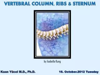
The Thoracic Cage • The skeleton of the chest-supports the thoracic cavity • Consists of: • thoracic vertebrae • ribs • sternum (breastbone) • Rib Cage - formed of ribs and sternum
Articulations of Ribs and Vertebrae • Functions • Protects organs of the thoracic cavity-heart, lungs, and thymus • Attaches muscles: • for respiration • of the vertebral column • of the pectoral girdle • of the upper limbs Figure 7–22b
The Ribs • Functions • are flexible • are mobile • can absorb shock • Rib movements (breathing): • affect width and depth of thoracic cage • changing its volume • Ribs (costae)-12 pairs of long, curved, flat bones extending from the thoracic vertebrae
Ribs 8–12 (false ribs): • do not attach directly to the sternum • Ribs 1–7 (true ribs) • vertebrosternal ribs • connected to the sternum by costal cartilages • Vertebrochondral ribs (ribs 8–10): • fuse together • merge with cartilage before reaching the sternum • Floating or vertebral ribs (ribs 11–12): • connect only to the vertebrae • have no connection with the sternum
Structures of the Ribs • The head (capitulum): • at the vertebral end of the rib • has superior and inferior articular facets • The neck: • the short area between the head and the tubercle • The tubercle (tuberculum): • a small dorsal elevation • has an auricular facet that contacts the facet of its thoracic vertebra (at T1–T10 only) • The tubercular body (shaft): • attaches muscles of the pectoral girdle and trunk • attaches to the intercostal muscles which move the ribs
The Sternum • 3 parts • 1-manubrium: • superior portion of sternum • broad, triangular shape • articulates with collarbones (clavicles) & cartilages of 1st rib pair • has a jugular notch between clavicular articulations • The sternum-a flat bone in the midline of the thoracic wall • 2-sternal body: • tongue-shaped • attaches to the manubrium & costal cartilages of ribs 2–7
3-xiphoid process: • smallest part of the sternum • attaches to the sternal body