Download
1 / 25
260 likes | 543 Vues
Applied Dentistry for Veterinary Technicians. Occlusion and Malocclusion. Occlusion. Orthodontics studies the way in which the teeth meet each other (occlude). Occlusion is defined as the normal position of the teeth when the jaws are closed.

E N D
Applied Dentistry for Veterinary Technicians Occlusion and Malocclusion
Occlusion Orthodontics studies the way in which the teeth meet each other (occlude). Occlusion is defined as the normal position of the teeth when the jaws are closed. In normal occlusion, the length and width of the jaws and the position of the teeth in the respective jaws are in harmony The development of the occlusion is determined primarily by genetic factors. The shape of the head affects the positioning of the teeth.
MesocephalicBreeds Well proportioned skull width and maxillary length; Dalmations, Labs & German shepherds.
Dolichocephalic Narrow skull and long maxilla; Sight hounds and Siamese cats.
Brachycephalic Breeds Wide skull with a short maxilla; Boxers, Bulldogs & Persian cats
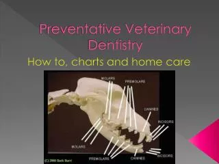
Normal “Scissor” Occlusion • In dogs, each lower canine tooth occludes in the interdental space between the upper third incisor and the upper canine tooth. In other words, the lower canine tooth occludes in front of the upper canine. • No incisor or canine tooth surface should touch. • The upper fourth premolar tooth overlaps the lower first molar, which together, constitute the carnassial teeth (shearing teeth). • In cats, normal occlusion is also a scissor occlusion with the upper incisors and canines slightly overlapping the lower incisors and canines slightly overlapping the lower incisors and canines. • The upper fourth premolar and lower molar are the feline carnassial (shearing teeth).
Normal scissors occlusion in a dog:Rostral view of incisors and canine teeth
Normal Scissors Occlusion: Lateral view of a dog skull. Premolar cusps interdigitate toward the opposing interdental space.
Malocclusion Malocclusion is an abnormality in the position of the teeth. It can occur in any of the three head shapes, but is more common in brachycephalic breeds. It is more common in dogs but also occurs in cats. Skeletal malocclusion results from jaw length and/or width discrepancy (usually inherited). Dental malocclusion results from tooth malpositioning. Four classes of malocclusions (Class I, II, III, and IV).
MandibularPrognathism “Underbite” or “Undershot jaw” Lower jaw is of normal length but the upper jaw is too short. These dogs will lose some of their self-cleaning ability and trap plaque and debris. Maxillary incisors can traumatize the mandible. Normally seen in brachycephalic breeds such as Bulldogs, Pekingese, Boston terriers, Pugs, and Persian cats.
Mandibular Prognathism Mandible is Longer than The maxilla
MandibularBrachygnathism “Overshot jaw,” “Overbite,” “Parrot mouth” Not as common as mandibularprognathism. Mandible is shorter than normal and dog has an “overbite.” Lack of self-cleaning ability and can create painful hard palate abrasion holes. Not an accepted standard in any breed. Often occurs in German Shepherd Dogs, Rottweilers, Collies, Standard Poodles and Dachshunds.
Upper jaw longer than the lower jaw. Mandible is longer than the maxilla.
Wry Mouth Each upper and lower right and left quadrant of the mouth is independent of the other, resulting in uneven growth which produces a wry occlusion (wry bite). In its mildest form, a one-sided prognathic or brachygnathic bite forms. In more severe cases, a crooked head and bite develop with a deviated midline. In some cases one side of the head may be smaller and the nose turned slightly to one side. A triangular opening (open bite) will also appear in the incisor area where the affected incisors do not meet. In severe cases, the tongue protrudes from the open bite.
Wry Mouth: One quadrant develops unevenly from the other quadrants
Anterior Crossbite Also called “Reverse Scissor.” Most common malocclusion in veterinary dentistry. Upper incisor teeth are caudal to the lower. May affect one, several, or all of the incisors. Thought to occur secondary to retained deciduous incisors. Can also be caused by tug-of-war games. Most common in medium and large breed dogs. May be a skeletal or dental malocclusion. Treatment consists of orthodontic movement or extraction of the abnormal teeth.
Posterior Crossbite Mandible is wider than the maxilla in the carnassial tooth area (area where the upper fourth premolar tooth overlaps the lower first premolar). Occurs occasionally in boxers, collies, and other dolichocephalic (long muzzled) breeds. More frequent professional prophylaxis will be needed for these pets.
Posterior Crossbite • Notice the upper fourth premolar tooth is positionedin an abnormal position inside the lower first molar tooth. Thiscondition is referred to as "posterior crossbite". This condition didnot require treatment as the pet had a comfortable and functionalocclusion (bite).
Retained Deciduous Teeth This is the most common dental problem seen in small animal practice. Mandibular canines, upper canines, and incisors. Interfere with normal eruption pathway of permanent teeth and are a reservoir for debris. Extraction is almost always necessary. Most common in small breed dogs. Note: All permanent incisors are positioned lingual to the deciduous teeth (babies in front). Permanent canine teeth will lie rostral to the deciduous teeth and appear whiter than the baby tooth (babies toward the back).
Polydontia Supernumerary teeth should not be confused with retained deciduous teeth. Radiographs aid in differentiation. Occurs in about 10% of dogs and rarely in cats. Commonly, the mandibular premolars and maxillary incisors are affected. Occasionally the extra teeth will erupt in an abnormal angle or be impacted. Unless extra tooth causes crowding, no treatment is necessary. If crowding is present, periodontal disease may result and spread to adjacent teeth. (Extraction necessary)
Oligodontia Also called hypodontia One or more teeth (usually incisors or premolars) do not form in the dental arcade. Many breeds are affected. In order to determine if tooth is unerrupted or hypodontia, an x-ray can be taken. If permanent tooth is absent, a baby tooth will often remain in the arch for months to years. No treatment; usually does not cause any problems.