Download

1 / 26

E N D
Myasthenia Gravis By Kim Anderson
Thymoma Facts A thymoma is a type of tumor or growth in the thymus gland. Thymic tumors are tumors of the thymus gland. • Physicians describe thymomas in terms of their degree of spread. Most thymomas have the potential to behave like a cancer and spread beyond the thymus, but many appear to behave in a benign fashion and are noninvasive. Less commonly, it appears to have spread beyond the thymus. People sometimes refer to such an invasive thymoma as malignant thymoma. When the pattern of spread is clearly typical for cancer, thymic carcinoma (cancer) is the term most often used. • The thymus gland is present in the front of the space between the upper lungs called the anterior mediastinum and behind the upper sternum. The thymus gland is larger during puberty but then normally becomes smaller in adulthood. • Thymomas most frequently appear in people in the fourth and fifth decades of life. • There are no known risk factors that predispose a person to developing a thymoma. • Up to half of thymomas are asymptomatic, meaning they do not produce any symptoms or signs and physicians diagnose them when they perform an imaging study of the chest for another reason. • Chest pain, shortness of breath, and cough are common symptoms that may be present when symptoms do occur. • Many patients with thymoma will have a so-called paraneoplastic syndrome. A paraneoplastic syndrome occurs preceding or concurrent with the discovery of the thymoma. These conditions accompany the cancer's development but are not a direct result of the disease as a lump or pain might be. They seem to be an indirect result of the cancer and may or may not improve with the treatment of the underlying disease. The most commonly associated condition with thymoma is myasthenia gravis, a disease of muscle. Twenty percent of patients with myasthenia gravis have a thymoma. • Thymomas are slow-growing tumors, and the prognosis is excellent when discovered in their early stages. • Surgical removal (surgical resection) is the mainstay of treatment. Chemotherapy, targeted therapy, and radiation therapy may be used in cases in which surgical treatment is not effective in removing the entire tumor or in particularly aggressive cases.
What is thymoma • A thymoma is a rare type of tumor of the thymus gland. The thymus is a gland located in the anterior mediastinum (the area between the two lungs and the sternum in the chest) that plays a critical role in the development of immune cells (lymphocytes) during childhood. The thymus gland enlarges during childhood, peaks in size at puberty (about 40 grams), and then begins to shrink. • Normally, a combination of lymphoid cells (immune cells or lymphocytes) and lining cells (epithelial cells) makes up the thymus. Thymoma is a type of tumor that originates from the epithelial or lining cells of the thymus. The term thymic neoplasms refers to tumors of the thymus, which consist of thymomas and thymic carcinomas. The term thymoma refers to tumors of the thymus that grow slowly and usually do not spread beyond the thymus. Thymic carcinomas are tumors of the thymus that grow aggressively and may metastasize to distant organs. Less than one person per 1.5 million people will develop a thymoma. This means about 400 people per year in the U.S. develop thymoma. Thymic carcinomas are very rare and make up only 0.06% of all thymic tumors.
What causes thymoma, and what are risk factors for thymoma? • The exact cause of thymomas is unknown. Thymomas are equally common in men and in women and are most frequently seen in the fourth and fifth decades of life. There are no known risk factors that predispose a person to developing thymoma.
What are signs and symptoms of thymoma? • Up to 50% of thymomas are asymptomatic, meaning they do not produce any symptoms or signs. Physicians may diagnose a thymoma when they perform an imaging study for another reason. In other cases, the tumor may cause symptoms related to the size of the tumor and the pressure it exerts on adjacent organs. • Chest pain, • shortness of breath, and • cough are common symptoms when symptoms do occur. • The following symptoms and signs are less common but may occur: • Fever • Night sweats • Weight loss • Some cases may spread to the lining of the lungs or heart or even to tissues outside the chest. Less than 7% of cases spread outside the chest cavity. Thymic carcinomas are more aggressive types of tumors than thymomas and are more likely to spread both locally and distantly (metastasize) and to cause symptoms.
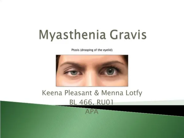
What other types of medical conditions are associated with thymoma? • A number of health conditions have been associated with thymoma. Medical conditions associated with cancers are paraneoplastic syndromes, and up to 50%-60% of patients with thymoma will have one of these related health conditions. The most commonly associated condition with thymoma is myasthenia gravis, an autoimmune disease of the nerve-muscle junction that can manifest as weakness, fatigue, double vision, ptosis (drooping eyelids), and problems with swallowing. • Other associated conditions include other autoimmune diseases including pure red cell aplasia (underproduction of red blood cells in the bone marrow).
What types of specialists treat thymomas? • Surgeons, including thoracic (chest) surgeons and surgical oncologists, typically treat thymoma. Medical oncologists and radiation oncologists may be involved in the treatment team if other treatments indicate an aggressive thymoma or thymic carcinoma.
How do health care professionals diagnose a thymoma? • If a thymoma is not causing symptoms, health care professionals may identify it incidentally, meaning it is found on an imaging test of the chest (for example, X-ray, computerized tomography or CT scan) that is performed for another reason. If symptoms are present, health care providers may carry out chest X-rays or other imaging studies, such as CT scans or magnetic resonance imaging (MRI) scans or the combination of PET and CT scans, to identify the source of the symptoms. • While a mass in the anterior mediastinum can be seen on imaging studies, the definitive diagnosis can only be established when the mass is either removed surgically and examined by a pathologist or when a biopsy (surgical removal of a small portion of tissue for diagnostic purposes) is taken. Microscopic examination of the tumor biopsy tissue is necessary to confirm the diagnosis of thymoma or thymic carcinoma. Health care professionals classify the appearance of the tissue itself under the microscope as type A, B, or C based on its characteristics. Type C thymoma is thymic carcinoma and is quite rare.
What are the stages of thymoma? • The stage of a tumor refers to the extent to which it has spread to other local organs and tissues or to other parts of the body. There are two commonly used staging systems for thymoma, both of which classify the tumors as stage 1 through stage 4, depending upon the extent of spread and the degree of tissue invasion. While there are some differences in the two methods used to stage thymomas, a stage 1 tumor represents an encapsulated tumor (surrounded by a capsular structure) that has not spread outside of the thymus gland. Stage 4 represents the opposite extreme, in which invasion and spread to distant organs has occurred. Locally recurrent thymoma refers to the situation in which a tumor returns in the same area after its surgical removal.
What are types of treatment options for thymoma? • Surgery is the primary treatment for thymoma. The success of the surgery depends upon the particular characteristics of the tumor and its precise location; tumors have a higher surgical cure rate if physicians can remove them completely. If all evidence of disease is unable to be removed and microscopic, or tumor remains after surgery, then radiation therapy, chemotherapy, and targeted therapy drugs have been used in addition to surgical resection.
What is the prognosis for thymoma? • The prognosis (outcome) for thymoma is dependent upon the stage of the tumor as well as the ability to remove the tumor by surgery. Thymic carcinomas tend to behave more aggressively and have a worse prognosis than thymomas. Thymomas tend to be slow-growing tumors, and the prognosis is good to excellent for those with stage 1 or stage 2 thymoma. It is hard to estimate exact survival statistics based on stage because of the low numbers of people diagnosed with this tumor. In a German study of patients whose thymomas were completely removed by surgery, only 3% of the tumors recurred. Even 83% of patients with stage 3 thymoma were alive 10 years after diagnosis. The 10-year survival rate for stage 4 thymoma is approximately 47%. Overall, a majority of thymoma patients will live at least five years, while fewer than half or those with thymic carcinoma are expected to live that long.
Is it possible to prevent a thymoma? • Because the cause of thymoma is unknown and no risk factors have been identified, prevention of thymoma is not possible.
Where in the Body Would a Thymoma Be Found? • A thymoma is a cancer that develops in the thymus gland. Located in the upper chest between the lungs, this gland plays an important role in maintaining the body’s immune system by developing T lymphocytes (T cells), a type of white blood cell that recognizes and attacks virus-infected cells. There are two kinds of thymomas – primary and secondary. Primary tumors develop in the thymus gland, while secondary tumors originate elsewhere in the body and metastasize (spread) to the thymus gland.
Thymoma in Myasthenia Gravis • Thymomas in myasthenia gravis (MG) are neoplasms derived from thymic epithelial cells, and are usually of the cortical subtype (WHO type B) [1]. 50% of thymoma patients develop MG (hereafter referred to as thymoma MG in this paper) [2, 3]. Cortical thymomas usually have some morphological similarities with thymic cortex; they share the capacity to propagate the maturation of immature naive CD4 T cells and export mature naive T cells into the periphery. Thymomas lacking this ability do not induce MG [4]. Thymomas with histological similarities to medullary thymic tissue or thymomas lacking developing T cells are seldom associated with MG [4]. Other thymoma characteristics that can cause reduced self-tolerance include defective epithelial expression of the autoimmune regulator (AIRE) gene and/or of major histocompatibility complex class II molecules, absence of myoid cells, failure to generate FOXP3(+) regulatory T cells, and genetic polymorphisms affecting T-cell signalling
Click to Histologically, thymomas are epithelial neoplastic cells surrounded by maturing T cells. The epithelial cells are capable of expressing epitopes cross-reactive with skeletal muscle proteins, such as acetylcholine receptor (AChR), titin, and ryanodine receptor (RyR) [6, 7]. The muscle-like epitopes are presented to T cells together with costimulatory molecules [7]. Autoreactive T cells specific for AChR and titin are found both in thymomas and in thymoma MG patients' sera [8]. Thymoma epithelial cells present AChR peptides to T-cell lines in thymoma MG patients, facilitating intrathymic immunization add text The patient's genetic profile and the thymic ability to export autoreactive T cells are equally important in developing MG. MG has a genetic association to HLA-DR3 or ancestral haplotype 8.1 in early-onset MG (MG onset before age 50 years) with thymic hyperplasia and several weaker associations to polymorphisms in immunoregulatory genes such as FcγR, TNF-α/β, GM-phenotypes, CTLA-4 [10], HLA, and PTPN22∗R620W [11]. The chance of having a thymoma increases with the number of thymoma-associated polymorphisms in an MG patient, indicating that thymoma MG is a polygenic disease and that thymoma patients with a particular genetic profile run higher risk of developing MG
Thymoma MG • MG is a neuromuscular junction disease characterized by muscular weakness and fatigability, caused in 85% of the cases by AChR antibodies [12]. When MG occurs together with a thymoma, MG is a paraneoplastic disease caused by the presence of the thymoma. Thymoma MG accounts for around 15% of all MG cases [13]. • The immune response against an epitope expressed on thymoma cells spills over to neuromuscular junction components sharing the same epitope [14]. In thymoma MG, epitopes are shared between the thymoma and muscle proteins.
Antibodies in Thymoma MG • AChR antibodies are the main cause of muscle weakness in thymoma MG [15]. Additional non-AChR muscle autoantibodies reacting with striated muscle titin and RyR antigens are found in up to 95% of MG patients with a thymoma and in 50% of late-onset MG patients (MG onset at age of 50 years or later) [16]. These antibodies are usually associated with more severe MG [13, 17–19]. Striational antibodies demonstrated in immunofluorescence are largely made up of titin antibodies [20]. • Titin is the largest known protein, with a molecular mass of 3000 kD stretching throughout the sarcomere, providing a direct link between mechanical muscle strain and muscle gene activation [21]. Myositis and myopathy with muscle atrophy are seen in some thymoma MG patients [22]. Sera from MG patients also induce degenerative changes in muscle cell cultures where both apoptosis and necrosis are implicated [23].
Click to adTheRyR is the calcium channel of the sarcoplasmic reticulum (SR). Upon opening, the RyR releases Ca2+ into the sarcoplasm resulting in muscle contraction. In vitro, RyR antibodies can inhibit Ca2+ release from the SR [24]. There is also a rat model with thymoma and MG with RyR antibodies but no AChR antibodies, indicating that RyR antibodies may cause MG symptoms irrespective of AChR antibodies [25]. There are also several reports of excitation-contraction coupling defects in thymoma MG [26].d text Click to add text
4. Recognizing the Clinical and Serological Pattern of Thymoma MG • MG patients with RyR antibodies are characterized by frequent involvement of bulbar, respiratory, and neck muscles at MG onset and a more severe disease. Neck weakness at MG onset is a distinctive feature of patients with RyR antibodies, while respiratory symptoms are also found in patients with titin antibodies with and without RyR antibodies. Limb involvement with few or no bulbar signs is typical at MG onset in RyR-antibody-negative MG [27]. Since many thymoma MG patients have RyR antibodies, neck weakness and nonlimb bulbar distribution of MG symptoms are initial characteristic features associated with thymoma MG. Such symptom distribution should always raise the suspicion about the presence of a thymoma in an MG patient. • Thymoma MG is equally frequent in males and females and occurs at any age with a peak onset around 50 years [28]. Thymoma MG and late-onset MG share similar serological profile with high prevalence of titin and RyR antibodies and lower AChR antibody concentrations compared to early-onset MG [29]. About 95% and 70% of thymoma MG patients have titin and RyR antibodies, respectively (Table 1). Around 58% and 14% of late onset MG patients have titin and RyR antibodies, respectively
Click to addLate MG onset age, similar serological profile, favorable pharmacological treatment response, severe MG, frequent use of immunosuppressive drugs, and the occurrence of MG related mortality are common features among thymoma MG and late-onset MG patients [29]. This profile differs from early-onset MG [30], that has higher AChR antibody concentrations, almost no titin or RyR antibodies, low need for immunosuppressive drugs, less severe MG, very low MG mortality rates, and a favorable thymectomy outcome [29]. Thymoma MG tends to be more severe than early-onset nonthymoma MG [29]. In one study, MG patients with thymoma or thymic atrophy (i.e., chiefly late-onset MG) had worse prognosis than MG patients with thymic hyperplasia (i.e., early-onset MG) [31]. The presence of a thymoma per se does not give a more severe MG. Thymoma MG patients and age-matched nonthymoma MG patients share similar MG long-term prognosis [19]. The presence of titin and RyR antibodies is associated with more severe disease in thymoma MG and in late-onset MG [29]. The AChR antibody serum concentration does not correlate with MG severity, mainly because of individual variations in AChR epitope specificity [32].
Verifying the Diagnosis of Thymoma MG • The diagnosis of MG is based on clinical disease history and typical clinical findings. MG can be confirmed pharmacologically by edrophonium (Tensilon) test which is positive in 90% of MG patients, giving an immediate but transitory improvement of MG signs [33]. The diagnosis of MG should be confirmed by the detection of AChR antibodies, present in most MG cases. These antibodies are present in virtually all patients with a thymoma [29]. In two thirds of MG patients, failure of neuromuscular transmission in leads to decremental response to repetitive nerve stimulation by electromyographical (EMG) examination [34]. Increased jitter on single-fiber EMG is even more sensitive than repetitive nerve stimulation when performed on affected muscles [34]. • In addition to MG, a thymoma should be demonstrated in order to fulfill the criteria of thymoma MG diagnosis. The diagnosis of a thymoma in MG is finally established by histopathological examination postsurgery. Titin and RyR antibodies and radiological examination of the anterior mediastinum share similar sensitivity for the presence of a thymoma in MG [29, 35, 36]. However, the presence of titin and RyR antibodies in a MG patient younger than 60 years strongly suggests a thymoma, while the absence of such antibodies at any age strongly excludes thymoma [13, 37]. Retesting for these antibodies and a new radiological examination should always be considered whenever clinical deterioration is seen over time, to minimize the risk of a previously undetected thymoma in a MG patient.
Surgical Treatment of Thymoma MG • When the diagnosis of a thymoma in a MG patient is established, the neoplasm should be removed surgically, and it is crucial to ensure radical excision of the neoplasm. Thymectomy can be performed transternally or through a video-assisted thoracoscopic approach, usually with similar outcome [38]. Radical excision of a thymoma does in most cases cure the thymic neoplasia, but patients will continue to suffer from MG after thymectomy, emphasizing the need of continuing followup and pharmacological treatment. When the thymoma invades the pleura or the pericardium, radical excision will not be possible and further oncological treatment is necessary. Presurgery plasmapheresis or intravenous infusion of immunoglobulin (iv-IgG) removes a great deal of circulating pathogenic antibodies [36]. In our department we give plasmapheresis or iv-IgG treatment to all patients with thymoma MG prior to thymectomy, to minimize the risk of postthymectomy MG exacerbation and myasthenic crisis. This practice varies however from department to another, and there is no consensus on this issue. Iv-IgG should be considered as first choice in patients at high risk of developing cardiopulmonary failure secondary to fluid overload caused by plasmapheresis [39]. MG outcome after thymectomy is generally less favorable in patients older than 45 years (i.e., mostly late-onset and thymoma MG patients)
7. Treatment of MG Crisis in Thymoma MG • Plasmapheresis and immunoglobulin treatments are also indicated in severe cases of thymoma MG regardless of thymectomy, such as in MG crisis and in severe MG cases with poor response to standard pharmacological treatment [41]. Parallel to plasmapheresis and immunoglobulin treatment, the pharmacological treatment should be intensified in these patients as explained in the next chapter.
Pharmacological Treatment of Thymoma MG • The first pharmacological choice in the treatment of thymoma MG is acetylcholinesterase inhibitors. The second choice is immunosuppressive drugs whenever additional pharmacological treatment is needed before or after thymectomy. Several immunosuppressive drugs are available, such as corticosteroids, azathioprine, cyclophosphamide, cyclosporine, methotrexate, mycophenolate mofetil, rituximab, and tacrolimus. Steroids such as prednisolone are frequently given on alternate days, by gradually raising the dose to 60–80 mg initially and then with slowly tapering to 20 mg or lower. If long-term treatment with steroids is regarded necessary, nonsteroid immunosuppressants such as azathioprine should be introduced in addition (usually 100–150 mg a day). While the steroid effect appears rapidly, the clinical effect of other immunosuppressants may take a few weeks to several months to develop [29]. Overall, about 80% of MG patients and 95% of thymoma MG patients need immunosuppressive drug treatment for more than one year
CliTacrolimus, which is an immunosuppressant and enhancer of RyR-related sarcoplasmic calcium release, may be especially beneficial in MG patients with RyR antibodies that in theory might block the RyR interfering with its function. Since most patients with thymoma MG have RyR antibodies, tacrolimus may act specifically in these patients. It may have a purely symptomatic effect in addition to its immunosuppressive impact [40]. Tacrolimus has demonstrated favorable effects in the treatment of MG, both as monotherapy and as add-on to prednisolone [43, 44]. Patients should undergo a thorough cardiological investigation prior to commencing tacrolimus treatment. Long-term observation of thymoma MG and age-matched nonthymoma MG patients showed no difference in MG severity over time, and both groups improved to the same degree after MG diagnosis as a result of pharmacological treatment and thymectomy. The need for immunosuppressive treatment in the two groups was similarly high. A thymoma that has been completely removed surgically does not necessarily mean worse MG prognosis in thymoma MG [19].