Download
1 / 1
20 likes | 188 Vues
Surface Plasmon Modes of a Silver Nanorod. Daniel C. Ralph, Cornell University, ECCS 0335765.

E N D
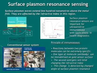
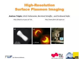
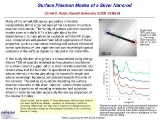
Surface Plasmon Modes of a Silver Nanorod Daniel C. Ralph, Cornell University, ECCS 0335765 Many of the remarkable optical properties of metallic nanoparticles (NPs) arise because of the excitation of surface plasmon resonances. The variety of surface plasmon resonant modes seen in metallic NPs is brought about by the dependence of surface plasmon excitation with the NP shape, size, composition and environment. Most applications of these properties, such as biochemical sensing and surface enhanced raman spectroscopy, are dependent on sub-wavelength spatial variations of the surface plasmons induced in the metal NPs.. In this study electron energy loss is characterized using energy filtered TEM of spatially resolved surface plasmon excitations on a silver nanorod supported on a silicon nitride substrate. Our results show that the excitation is quantized as resonant modes whose intensity maxima vary along the nanorod’s length and whose wavelength becomes compressed towards the ends of the nanorod. Theoretical calculations modeling the surface plasmon response of the silver nanorod - silicon nitride system show the importance of including retardation and substrate effects in order to describe accurately the energy dispersion of the resonant modes. Bright-field (BF) zero-loss image of a 666 ± 3 nm long silver nanorod, with 47 ± 3nm diameter. Olivia Nicoletti, Martijn Wubs, N. Asger Mortensen, Wilfried Sigle, Peter A. van Aken, and Paul A. Midgley, University of Cambridge, Technical University of Denmark, and Max Planck Institute for Intelligent Systems Silver nanorods synthesized at Washington University in St. Louis Nano Research Facility Electron energy-loss spectra (unprocessed) acquired at the positions marked in the bright-field image of matching color. Spectra have been obtained using a selected-area approach and shown displaced on the intensity axis for clarity.