Download
1 / 94
1.95k likes | 3.94k Vues
Complications of Sinusitis. Dr. Vishal Sharma. Definition. 1. Adverse progression of infection beyond muco-periosteal lining of para nasal sinuses to involve bone & neighboring structures (orbit, intra-cranial cavity, dentition)

E N D
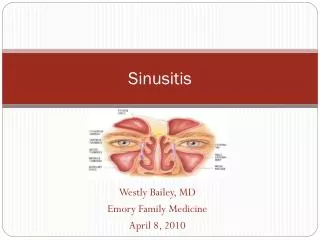
Complications of Sinusitis Dr. Vishal Sharma
Definition 1. Adverse progression of infection beyond muco-periosteal lining of para nasal sinuses to involve bone & neighboring structures (orbit, intra-cranial cavity, dentition) 2. Compromise in function of any part of body due to sinusitis
Etiology 1. Weak immune response of host: young children & immuno-compromised adults 2. Inadequate or inefficient treatment 3. Infection by high virulence organisms 4. Abnormalities of muco-cilliary clearance 5. Persistent allergy & blockade of sinus ostia
Routes of infection 1. Via thin bones: lamina papyracea 2. Through natural suture lines 3. Through natural canal: infra-orbital canal 4. Retrograde thrombophlebitis: diploic vein of Breschet 5. Closely related roots of upper 2nd premolar & 1st molar teeth 6. Peri-arteriolar space of Virchow Robin
Common pathogens • Staphylococcus aureus • Streptococcus pnemoniae • Haemophilus influenzae • Moraxella catarrhalis • Anaerobes: Bacteroides • Aspergillus • Rhizopus
Classification A. AcuteB. Chronic 1. Local Mucocele (?) Orbital Pyocele (?) Intra-cranial C. Associated diseases Bony Otitis media Dental Adeno-tonsillitis 2. Distant Bronchiectasis Toxic shock Atrophic rhinitis syndrome Nasal polyp
Orbital Complications 1. Pre-septal cellulitis 2. Orbital cellulitis without abscess 3. Orbital cellulitis with extra-periosteal abscess 4. Orbital cellulitis with intra-periosteal abscess 5. Cavernous sinus thrombosis ?: intracranial 6. Orbital apex syndrome
Intracranial Complications 1. Meningitis 2. Encephalitis 3. Extra-dural abscess 4. Sub-dural abscess 5. Intra-cerebral abscess 6. Cavernous sinus thrombosis 7. Sagittal sinus thrombosis
Other local complications Bony 1. Osteitis 2. Osteomyelitis (Pott’s puffy tumour) Dental 1. Dental abscess 2. Oro-antral fistula
Introduction • Commonest complication of sinusitis • Young people at high risk: 85% < 20 yrs age • Ethmoid sinus most commonly implicated Frontal Sphenoid Maxillary • Left orbit more commonly involved
Pre-septal cellulitis • Infection external to peri-orbital septum • Edema of eyelid:upper lid = frontal sinusitis lower lid = maxillary sinusitis both lids = ethmoid sinusitis • No erythema / tenderness / proptosis / extra-ocular movement restriction / vision change
Orbital Cellulitis • Infection inside peri-orbital septum • Diffuse peri-orbital edema • Mild proptosis present • Minimal or no restriction of extra-ocular movement • No change in vision
Extra-periosteal abscess • Localized extra-periosteal pus collection • Mild proptosis present • Mild restriction of extra-ocular movement • Mild vision loss • Color vision affected first: Red = brown Blue = black
Intra-periosteal orbital abscess • Mild chemosis • Proptosis: severe, asymmetric, quadrantic Frontal sinusitis = down + forward + lateral Ethmoid sinusitis = forward + lateral Maxillary sinusitis = up + forward • Concurrent, complete, ophthalmoplegia • Severe vision loss
Cavernous Sinus Thrombosis • Rapid onset, hectic fever • Bilateral orbital pain + severe chemosis • Bilateral absent pupillary reflex • Bilateral symmetrical axial proptosis • Sequential ophthalmoplegia (VI III IV) • Papilloedema + loss of vision • Painful paraesthesia of V1, V2
C.T. with venogram Absence of contrast in cavernous sinuses
Orbital apex syndrome • Frontal headache + deep orbital pain • Optic nerve involvement (vision loss) • Paralysis of abducens nerve • Paralysis of oculomotor nerve • Paralysis of trochlear nerve • Painful paraesthesia of V1, V2
Evaluation of orbital complication 1. Eye examination: Ophthalmology consultation • Edema of eyelids • Displacement of eyeball • Ocular movement • Visual acuity • Fundoscopy for papilledema 2. CT scan PNS (including orbit): coronal & axial
Medical Treatment 1. Broad spectrum, high dose IV antibiotics (Ceftriaxone + Metronidazole) 2. NSAIDs 3. Topical / oral decongestants 4. Mucolytics: Bromhexine, Ambroxol 5. Nasal saline irrigation
Surgical Treatment For sinusitis: 1. Frontal trephination 2. External fronto-ethmoidectomy (Lynch Howarth) 3. Functional Endoscopic Sinus Surgery For orbital complication: 1. Sub-periosteal abscess drainage 2. Orbital decompression
Sub-periosteal abscess drainage • Incision made b/w caruncle (C) & semilunar fold (S) • Tissue b/w caruncle & semilunar fold incised with tenotomy scissors • Periosteum (P) incised & elevated with Freer elevator until abscess (A) is found & drained
Indications for orbital decompression • No improvement in orbital symptoms in 24- 48 hours of treatment • CT scan evidence of orbital abscess • Visual acuity of 20 / 60 or worse
Techniques of decompression 1. Patterson’s trans-orbital approach 2. Endoscopic intra-nasal approach 3. Trans-antral approach 4. Combined intra-nasal & trans-antral approach • Medial wall + floor of orbit removed • Removal of 1 wall = 2 - 3 mm decompression • Removal of 2 walls = 4 - 7 mm decompression
Result of orbital complications • Exposure keratitis • Uveitis • Choroiditis • Ophthalmoplegia • Glaucoma • Permanent vision loss
Introduction • 2nd commonest complication of sinusitis • Most common in adolescents & young adults (diploic venous system at peak vascularity) • Frontal sinus most commonly implicated Ethmoid Sphenoid Maxillary • Commonest route of spread = retrograde thrombophlibitis via Diploic vein of Breschet