Download
1 / 44
440 likes | 469 Vues
Unit 1 Chapter 1 Bacterial Cell Structure. CLS 3303 Clinical Microbiology. Taxonomy . Defined as the orderly classification & grouping of organisms into categories Kingdom, Division, Class, Order, Family, Tribe, Genus and Species ( these are the formal levels of classification)

E N D
Unit 1 Chapter 1Bacterial Cell Structure CLS 3303 Clinical Microbiology
Taxonomy • Defined as the orderly classification & grouping of organisms into categories • Kingdom, Division, Class, Order, Family, Tribe, Genus and Species ( these are the formal levels of classification) • Family = “Clan”; has “–aceae” ending • Genus = “Human last name” • Species = “Human first name” • When in print, genus and species are italicized. (Staphylococcus aureus) • , When written genus and species are underlined. (Staphylococcus aureus) • To abbreviate organism names: use first letter capitalized of the genus followed by a period and the species epithet. ( i.e S. aureus)
Nomenclature • Staphylococcus sp. is used when referring to the genus as a whole when the species is not identified. • “sp.” – singular (Staphylococcus sp.) • “spp.” – plural (Staphylococcus spp.)
Classification by Cellular Type • Bacteria Identification – test each bacterial culture for a variety of metabolic characteristics and compare the results with known results. • All organisms are either “prokaryotes”, “eukaryotes”, or “archaeobacteria”
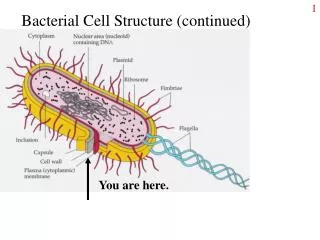
Classification by Cellular Type: Prokaryotes • PROKARYOTES - bacteria • Do not have a membrane-bound nucleus • DNA is a single circular chromosome and RNA are free in the cytoplasm • Have both cell (plasma) membrane AND cell wall. • Have no mitochondria, endoplasmic reticulum (ER) or Golgi bodies
Classification by Cellular Type: Eukaryotes • EUKARYOTES - fungi, algae, protozoa, animal cells, and plant cells • Cells have nuclei that contains DNA and are complex • Most cells do NOT have a cell wall (Fungi have cell walls made chitin)
Classification by Cellular Type: Archaeobacteria • Resembles eukaryotes • Found in microorganisms that grow under extreme environmental conditions • Cell wall lacks peptidoglycan • See chart on page 5 for comparisons of Prokaryotes and Eurkaryotes
Gram Positive (G+) Cell Wall • Very thick protective peptidoglycan layer • Many G+ antibiotics act by preventing synthesis of peptidoglycan • Consists of cross-linked chains of glycan • Also contain teichoic acid and lipoteichoic acid • Unique structure makes these bacteria G+ by protecting against the decolorizing step in Gram staining
Gram Negative (G-) Cell Wall • Two layers; outer is much thinner than G+ cell walls • Outer wall contains several molecules, including Lipid A which is responsible for producing fever and shock in infections with G- bacteria • The thin walls allow the decolorizer to enter the cell and take out the crystal violet stain.
(G+) and (G-) Microorganisms • G+ cocci in clusters→ • G- bacilli (rods)→ • When identifying bacteria, remember that rods can sometimes be short and look like cocci, but cocci do not look like rods
Acid Fast Cell Wall • Mainly Mycobacteria and Nocardia • Have a G+ cell wall structure but also a waxy layer of glycolipids and fatty acids (mycolic acid). It is hydrophobic and affects permeability • Waxy layer makes them difficult to gram stain (More than 60% of the cell wall is lipid) • Cannot be decolorized by acid-alcohol, hence the name “acid fast” • Bacteria is pink • Background is green or blue
Absence of Cell Wall • Mainly Mycoplasma and Ureaplasma • Lack of cell wall results in a variety of shapes microscopically • Contain sterols in cell membrane
Surface Polymers: Slime Layers • Some bacteria produce slime layers • Made of polysaccharides • Inhibit phagocytosis and also help to attach to the host.
Surface Polymers: Capsule • Some bacteria produce a capsule • Protect the bacteria from phagocytosis • Capsule usually does not stain, but can appear as a clear area (halo-like)
Cell Appendages • Flagella – exterior protein filaments that rotate and cause bacteria to be motile • Polar • Extend from one end • Can occur singly or in multiple tufts • Peritrichous • Flagella found on all sides of bacteria • Pili (fimbriae) – hairlike projections that aid in attachment to surfaces
Bacterial Morphology • Microscopic Shapes • Cocci (spherical) • Bacilli (rod-shaped) • Spirochetes (helical) • Groupings • Singly • Pairs • Clusters • Chains • Palisading
Bacterial Morphology (cont’d) • Size and length • Short • Long • Filamentous: • Fusiform: bacilli with tapered, pointed ends • Curved • Pleomorphic: variance in size & shape within a pure culture
Other Common Bacterial Stains: Acridine Orange (fluorochrome dye) • Stains nucleic acid of both G+ and G- bacteria, either living or dead; used to locate bacteria in blood cultures and other specimens where background material obscures gram stains
Other Common Bacterial Stains: Methylene Blue • Stain for Corynebacterium diphtheriae to show metachromatic granules and as counter-stain in acid-fast stain procedures
Other Common Bacterial Stains: Lactophenol Cotton Blue – fungal stain
Other Common Bacterial Stains:Calcuflour White – fungal stain • A fluorochrome that binds to chitin in fungal cell walls • Apple-green or blue-white with a fluorescent microscope
Other Common Bacterial Stains: India Ink • Negative stain for capsules, surrounds certain yeasts
Other Common Bacterial Stains:Endospore stain • Heat is used to help the primary stain (Malachite green) into the spore. The spore stains green • The counter stain, (safranin) stains the rest of the organism
Microbial Growth and Nutrition Needs • Source of carbon for making cellular constituents • Source of nitrogen for making proteins • Source of energy (ATP) for cellular functions • Smaller amounts of other molecules
Nutritional Requirements for Growth • Autotrophs (lithotrophs) • Able to grow simply, using only CO2, water and inorganic salts • Obtain energy via photosynthesis or oxidation of inorganic compounds • Occur in nature and do not normally cause disease
Nutritional Requirements for Growth • Heterotrophic • Require more complex substances for growth • Require an organic source of carbon and obtain energy by oxidizing or fermenting organic substances • All human bacteria fall in this category • Within this group, nutritional needs vary greatly
Types of Growth Media • Minimal medium – simple; not usually used in diagnostic clinical microbiology • Nutrient medium – made of extracts of meat or soy beans • Enriched medium – nutrient medium with extra growth factors, such as blood • Selective medium – contains additives that inhibit the growth of some bacteria while allowing others to grow • Differential medium – contains additives that allow visualization of metabolic differences in bacteria • Transport medium – holding medium to preserve those bacteria present but does not allow multiplication
Environmental Factors Influencing Growth • pH – most media is between 7.0 and 7.5 • Temperature – most pathogens grow at body temperature; grown at 35° C in the lab
Environmental Factors Influencing Growth • Gaseous composition • Obligate aerobes – require oxygen • Obligate anaerobes – cannot grow in the presence of oxygen • Facultative anaerobes – can grow with or without oxygen • Capnophilic – grow better with extra CO2 (5 -10%) • Microaerophilic- grow better in low oxygen environments ( about 20%) • Campylobacter spp. require 5 – 6% oxygen
Bacterial Growth • Reproduce by binary fission • Can be fast (as little as 20 minutes for E. coli) or slow (as long as 24 hours for M. tuberculosis)
Determination of the Number of bacterial cells • Direct counting under microscope: estimate the number of bacteria in a specimen. Does not distinguish live or dead cells • Direct plate count: grown from dilutions of broth cultures. Counts viable cells only. Colony Forming Units (CFU/mL) • Density measurement: (turbidity) bacterial broth culture in log phase
Bacterial Biochemistry and Metabolism • Metabolic reactions cause production of energy in form of ATP • Identification systems analyze unknown specimens for: • Utilization of variety of substances as a source of carbon • Production of specific end products from various substrates • Production of acid or alkaline pH in the test medium
Fermentation • Anaerobic process in obligate and facultative anaerobes • The electron acceptor is an organic compound • Does NOT require oxygen
Oxidation (Respiration) • More efficient energy-generating process • Molecular oxygen is the final electron acceptor • Aerobic process in obligate aerobes and facultative anaerobes
Metabolic Pathways • Main one is Embden-Meyerhoff • Convert glucose to pyruvic acid, a key intermediate • Generates energy in the form of ATP
Metabolic Pathways • From pyruvic acid: • Alcoholic fermentation (ethanol) (ex: yeast) • Homolactic acid fermentation (lactic acid) )ex: strep) • Heterolactic acid fermentation (lactic acid, CO2, alcohols, formic and acetic acids • Propionic acid • Mixed acid fermentation (lactic, acetic, succinic, and formic) (ex: e-coli and salmonells) • Butanediol fermentation: (ex: Kleb, enterobacter & serratia) • Butyric acid fermentation: (ex: obligate anaerobe)
Metabolic Pathways • Main oxidative pathway is the Krebs Cycle, resulting in acid and CO2 • Carbohydrate Utilization & Lactose Fermentation • “Sugars” = carbohydrates • Lactose fermentation – key component in identification schemes • Lactose is converted to glucose, so ALL lactose fermenters also ferment glucose
Genetic Elements and Alterations • Plasmid • Extra piece of DNA • Code for antibiotic resistance and other virulence factors are often found on plasmids • Sometimes passed from one bacterial species to another. This is how resistance is acquired.
Genetic Elements and Alterations • Mutations • “They don’t always read the book” • Changes that occur in the DNA code • Results in changes in the coded protein or in the prevention of its synthesis
References • http://media.photobucket.com/image/micro/lovitex2000/Micro%20biology%20lab/b1cf.jpg?o=81 • http://nhscience.lonestar.edu/biol/wellmeyer/bacteria/capsules3.jpg • http://www.iccb.state.il.us/pt3/images/sci/mod11/bacillus_subtilis.jpg