Download
1 / 74
760 likes | 973 Vues
Compartment Syndromes. T. Toan Le, MD and Sameh Arebi, MD Original Author: Robert M. Harris, MD; Created March 2004 New Authors: T. Toan Le, MD and Sameh Arebi, MD; Revised December 2005; Revised May 2011 – Michael Sirkin. Today. What is it Pathophysiology Diagnosis Treatment.

E N D
Compartment Syndromes T. Toan Le, MD and Sameh Arebi, MD Original Author: Robert M. Harris, MD; Created March 2004 New Authors: T. Toan Le, MD and Sameh Arebi, MD; Revised December 2005; Revised May 2011 – Michael Sirkin
Today • What is it • Pathophysiology • Diagnosis • Treatment
Increase in hydrostatic pressure in closed osteofascial space resulting in decreased perfusion of muscle and nerves within compartment
RAISED PRESSURE WITHIN A CLOSED SPACE with a potential to cause irreversible damage to the contents of the closed space
Richard Von Volkmann, 1881 • “For many years I have noted on occasion, following the use of bandages too tightly applied, the occurrence of paralysis and contraction of the limb, NOT … due to theparalysis of the nerve by pressure, but as a quick and massive disintegration of the contractile substance and the effect of the ensuing reaction and degeneration.”
Definition • Symptoms resulting from increased pressure within a limited space • compromising • circulation • function
Pathophysiology • Local Blood Flow is reduced as a consequence: LBF=Pa-Pv / R (A-V Gradient)
Pathophysiology • A continuous increase in pressure within a compartment occurs until the low intramuscular arteriolar pressure is exceeded and blood cannot enter the capillaries
Pathophysiology • Increased compartment pressure Increased venous pressure Decreased blood flow Decreases perfusion
Pathophysiology • Autoregulatory mechanisms may compensate: • Decrease in peripheral vascular resistance • Increased extraction of oxygen • As system becomes overwhelmed: • Critical closing pressure is reached • Oxygen perfusion of muscles and nerves decreases
Muscle Ischemia • 4 hours - reversible damage • 8 hours - irreversible changes • 4-8 hours - variable Hargens JBJS 1981
Muscle Ischemia • Myoglobinuria after 4 hours • Renal failure • Maintain a high urinary output • Alkalinize the urine • Cell death initiates a “vicious cycle” • increase capillary permeability • increased muscle swelling
Increased muscle swelling Increased permeability Increased compartment pressure
Increased pressure Increased venous pressure Decreased blood flow Decreases perfusion
Repetitive Cycle Increased muscle swelling Increased permeability Increased compartment pressure
Nerve Ischemia • 1 hour - normal conduction • 1- 4 hours - neuropraxic damage reversible • 8 hours - axonotmesis and irreversible change Hargens et al. JBJS 1979
Pathophysiology: • CAUSES: • Increased Volume - internal : hemmorhage, fractures, swelling from traumatized tissue, increased fluid secondary to burns, post-ischemic swelling • Decreased volume - external: tight casts, dressings • Most common cause of hemmorhage into a compartment: fractures of the tibia, elbow, forearm or femur
Etiology • Fractures • Soft Tissue Injury (Crush) • Arterial Injury • Post-ischemic swelling • Reperfusion injury • Drug Overdose (limb compression) • Burns
Pathophysiology: Most common cause of compartment syndrome is muscle injury that leads to edema
Arterial Injuries • Secondary to revascularization: • Ischemia causes damage to cellular basement membrane that results in edema • With reestablishment of flow, fluid leaks into the compartment increasing the pressure
Diagnosis • Clinical diagnosis • High index of suspicion • Syndrome • History • Physical Exam
Difficult Diagnosis • Classic signs of the 5 P’s - ARE NOT RELIABLE: • pain • pallor • paralysis • pulselessness • paresthesias • These are signs of an ESTABLISHED compartment syndrome where ischemic injury has already taken place • These signs may be present in the absence of compartment syndrome.
Diagnosis • Pain • Compartment pressure • Confirmatory test • Don’t just measure
Diagnosis • Palpable pulses are usually present in acute compartment syndromes unless an arterial injury occurs • Sensory changes and paralysis do not occur until ischemia has been present for about 1 hour or more
Diagnosis • The most important symptom of an impending compartment syndrome is PAIN DISPROPORTIONATE TO THAT EXPECTED FOR THE INJURY
Signs & Symptoms • Pain • Passive muscle stretching • Out of proportion • Progressive • Not relieved by immobilization
Signs & Symptoms • Pain • May be worse with elevation • Patient will not initiate motion on own • Be careful with coexisting nerve injury
Signs & Symptoms • Parasthesia • Secondary to nerve ischemia • Must be differentiated from nerve injury
Signs & Symptoms • Paralysis (Weakness) • Ischemic muscles lose function
Signs & Symptoms • Tense compartment on palpation • Elevated compartment pressure
Tissue Pressure • Normal tissue pressure • 0-4 mm Hg • 8-10 with exertion • Absolute pressure theory • 30 mm Hg - Mubarak • 45 mm Hg - Matsen • Pressure gradient theory • < 20 mm Hg of diastolic pressure – Whitesides • < 30 mm Hg of diastolic pressure McQueen, et al
Tissue-Pressure: Principles • Originally, fasciotomies for tissue-pressures greater-than 30mmHg • Whitesides et al in 1975 was the first to suggest that the significance of tissue pressures was in their relation to diastolic blood pressure. • McQueen et al: absolute compartment pressure is an UNRELIABLE indication for the need for fasciotomies. BUT, pressures within 30mmHg of DP indicate compartment syndrome
Tissue-Pressure: Principles • Heckman et al demonstrated that pressure within a given compartment is not uniform • They found tissue pressures to be highest at the site or within 5cm of the injury • 3 of their 5 patients requiring fasciotomies had sub-critical pressure values 5cm from the site of highest pressure
High energy fractures • Severe comminution • Joint extension • Segmental injuries • Widely displaced • Bilateral • Floating knee • Open fractures
Impaired Sensorium • Alcohol • Drug • Decreased GCS • Unconscious • Chemically unconscious • Neurologic deficit • Cognitively challenged
Diagnosis • The presence of an open fracture does NOT rule out the presence of a compartment syndrome • 6-9% of open tibial fractures are associated with compartment syndromes • McQueen et al found no significant differences in compartment pressures between open and closed tibial fractures • No significant difference in pressures between tibial fractures treated with IM Nails and those treated with Ex-Fix
Criteria-Compartment Pressure • Accurately examine • Difference < 30mm Hg • Impaired • Absolute > than 30mm Hg
Needle Infusion Technique-Historical • Needle inserted into muscle, tube with air/saline interval kept at this height, manometer indicates pressure • Air injected by syringe via 3-way stopcock • When the pressure of the injected air exceeds the compartment pressure pressure, the saline interval moves in the tube • AT this point, the second person reads the pressure from the manometer NEED 2 PEOPLE ! saline
Pressure Measurement • Infusion • manometer • saline • 3-way stopcock (Whitesides, CORR 1975) • Catheter • wick • slit catheter • Arterial line • 16 - 18 ga. Needle (5-19 mm Hg higher) • transducer • monitor • Stryker device • Side port needle
Pressure Measurement • Needle • 18 gauge • Side ported • Catheter • wick • slit • Performed within 5 cm of the injury if possible-Whitesides, Heckman Side port
Unit and needle set • Assemble unit and prime • Hold at angle to measure • Zero machine • Test each of 4 compartments • Keep calf off of bed
Most Common Locations • Leg: deep posterior and the anteriorcompartments • Forearm: volar compartment, especially in the deep flexor area
Pressure • Deeper muscles are initially involved • Distance from fracture affects pressure Heckmen et al. JBJS 1994
Compartments • Anterior • Lateral • Posterior • Deep • Superficial
Compartments • Anterior • Lateral • Posterior • Deep • Superficial TA EDL EHL Peroneus TP FDL FHL Soleus Gastroc
Treatment • Remove restricting bandages • Serial exams • When diagnosis made • Immediate surgery • 4 compartment fasciotomy
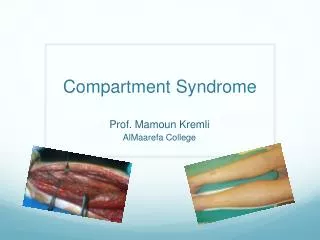
Treatment THE ONLY EFFECTIVE WAY TO DECOMPRESS AN ACUTE COMPARTMENT SYNDROME IS BY SURGICAL FASCIOTOMY!!! (unless missed compartment syndrome)
Treatment • Fasciotomy • One incision • With or without Fibulectomy • Two incisions • All 4 compartments must be released • Not selective