Download
1 / 24
270 likes | 590 Vues
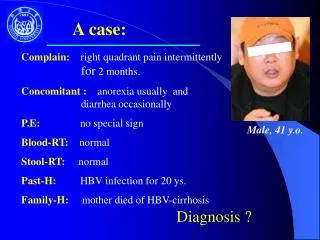
HKCEM College Tutorial. A Lady with Right Upper Quadrant P ain. Author Dr. LEE KF, Dr. TANG CO, Dr. TAM MK Revised by DR CHAN CHI MING May 2013. Triage Notes. F/75 c/o: RUQ pain for 1 day PMH: DM, IHD, old PTB BP 172/78 P 96/min T 37.8 ºC. Triage Cat III.

E N D
HKCEM College Tutorial A Lady with Right Upper Quadrant Pain Author Dr. LEE KF, Dr. TANG CO, Dr. TAM MK Revised by DR CHAN CHI MING May 2013
Triage Notes • F/75 • c/o: RUQ pain for 1 day • PMH: DM, IHD, old PTB • BP 172/78 • P 96/min • T 37.8 ºC Triage Cat III
What are your differential diagnosis ? • Cholecystitis • Cholangitis • Liver abscess • PPU • Basal pneumonia • AMI… What would you like to ask in the history ?
History • Fever & system review • CVS ischemia • Resp cough… • GU renal colic • PMH • Current med • Drug allergy • Pain = PQRST • Place RUQ • Quality constant • Radiate to scapula • Severity e.g. pain score • Time for one day • GI symptoms • N, V, D, • Jaundice, tea color urine
Physical Findings • A fat lady with T 37.8°C • No pallor or jaundice • A palpable globular shape mass at RUQ of abdomen • Murphy’s sign positive • Other systems essentially normal
What is Murphy’s sign ? • Murphy’s sign is a reliable sign of acute cholecystitis • The patient is asked to take a deep breath while the examiner palpates the gallbladder region • The breath catches at the zenith of inspiration as the inflamed gallbladder moves down into contact with the examining hand • Elicit tenderness Positive Demonstration
What is Courvoisier’s law ? • Courvoisier’s law states that • if the gallbladder is palpable in the presence of jaundice, the jaundice is unlikely to be due to stone
What is Charcot’s triad ? = RUQ pain + pyrexia + jaundice • It suggests a diagnosis of cholangitis
What is the differential diagnosis of RUQ mass ? • Gallbladder • Liver • Colon
X-ray Blood ECG Ultrasound What investigations would you order ? Progress
What are the findings ? X-ray • RUQ opacity likely gallstone • No pneumobilia • No dilated bowel • Clear lung fields • No free gas
X-ray CXR (erect film) • to look for free gas under diaphragm, which may be caused by PPU or perforated gallbladder in case of acute cholecystitis AXR • to look for calcified lesion suggesting gallstone • Also look for sign of IO (gall stone ileus in case of acute cholecystitis)
Gallstone • Only 15% of stones contain enough calcium to be seen on a plain film • Ultrasound is a much more sensitive test for gallstone
Blood CBC • Hb 12 g/dL • WCC 12 x10^9 /L RFT • Urea 6.2 mmol/L • Cr 76 umol/L LFT • Bili 12 umol/L • ALT 57 U/L • ALP 40 U/L Amylase 89 U/L Tn-I < 0.03 mmol/L Glucose / H’stix 13.7 mmol/L
ECG AF with ventricular rate of about 90/min no acute ischemic changes
What are the findings ? Ultrasound Thickened GB wall & peri-cholecystic fluid Distended Gallbladder Gallstone with echogenic foci & posterior acoustic shadow
What are the findings ? Ultrasound • Echogenic foci with posterior acoustic shadowing suggesting gallstones • Distended gallbladder • Gallbladder wall thickened • Peri-cholecystic fluid • Positive sonographic Murphy’s sign • Not demonstrate in the picture Compatible with acute cholecystitis • Also look for: • Ductal dilatation (CBD & intrahepatic) to exclude cholangitis • Liver tumor as hepatitis B is prevalent in our locality • Sub-hepatic fluid collection (abscess) US demonstration
Ultrasound • What is positive sonographic Murphy’s sign? • What are the sonographic diagnostic criteria for acute cholecystitis? Positive sonographic Murphy’s sign —tenderness elicited by pressing the gallbladder with the US probe
What are the sonographic diagnostic criteria for acute cholecystitis ? Major diagnostic criteria: • Positive sonographic Murphy’s sign • Loss of definition of gallbladder margins • Thickening of the gallbladder wall > 3 mm • Linear or irregular hypoechoic areas within the wall • Peri-cholecysticfluid • Intramural gas Minor criteria: • Distended gallbladder • Presence of calculi or sludge (5% of cases are not associated with gallstones) (If patient does not have ascites, chronic liver disease & right heart failure) (Indicative of impending perforation) Details
Treatment Progress • Resuscitate if necessary • Analgesia • Admit to surgical unit for further management
Treatment after admission Progress • Confirm the diagnosis by blood tests, formal USG (by radiologist) • Conservative treatment (include: NPO, IV fluid, antibiotics, NG suction if appropriate) • 90% of cases will settle • Close monitor for complications: • sepsis, empyema, gangrene & perforation • Surgical treatment • laparoscopic or open cholecystectomy • If medically not fit for GA, may perform percutaneous cholecystostomy
The End Any Questions ?