Download
1 / 84
840 likes | 951 Vues
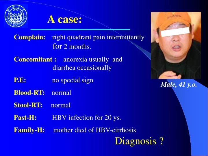
A case:. Complain: right quadrant pain intermittently for 2 months. Concomitant : anorexia usually and diarrhea occasionally P.E: no special sign Blood-RT: normal Stool-RT: normal Past-H: HBV infection for 20 ys.

E N D
A case: Complain: right quadrant pain intermittently for 2 months. Concomitant : anorexia usually and diarrhea occasionally P.E: no special sign Blood-RT: normal Stool-RT: normal Past-H: HBV infection for 20 ys. Family-H: mother died of HBV-cirrhosis Male, 41 y.o. Diagnosis ?
Hepato-Cellular Carcinoma (HCC) Chang-qing Yang, MD & PhD Tongji Hospital of Tongji University 同济大学附属同济医院 杨长青
Special Terms • Hepatocellular Carcinoma (HCC) 肝细胞癌 • Aflatoxin β1 黄曲霉素B1 • -Fetoprotein (AFP) 甲胎球蛋白 • -Glutamyl Transferase ( -GT)-谷氨酰转移酶 • -L-Fucosidase (ALF)-盐藻糖苷酶 • Des- -Carboxy Prothrombin (D -- CP)脱羧基凝血酶原 • Hepatomegaly肝肿大 • TACE (transarterial chemoembolization ) 介入化疗
Background: HCC • most common solid organ tumor worldwide • incidence is rising: USA 2.4 /100,000 China 20.4 /100,000 South Africa 60 / 100,000 • in China, marginal waters > hinterland eastern > western one million died annually, Top 2 in tumors
Sex distribution • Male :Female = 3~5 :1 in China • Almost equal in developed countries
Age distribution • Middle aged man in predominance (43.7 y) • It is rare in children • It rises progressively with age, although it tends to level off in the oldest age group
Etiology & Pathogenesis • Four major causal associations of HCC • The etiologic agents differs with different area
Risk factors Major • Chronic HBV infection • Chronic HCV infection • Aflatoxin β 1 • Liver fibrosis & Cirrhosis
Hepatitis B virus • Chronic infection with HBV may cause as much as 80% of human HCC • It is closest in ethnic Chinese and black Africans • Early infection carries a greater risk
HBV & HCC
Hepatitis B Virus • HBV DNA could be integrated into cellular DNA in about 95% of patients with HBV-related tumors • The site of integration is random • Integration perturbing the function of cellular oncogenes or tumor suppressor genes, which may contribute to hepatocellular carcinogenesis
Hepatitis C Virus • HCV is carcinogenic in human • In Japan, Italy, Spain, chronic HCV infection is the major risk factor for hepatocellular carcinoma • A far smaller percentage of ethnic Chinese and black African have HCV-induced tumors • HCV does not integrate into host DNA, its mechanism differs from that of HBV
HCV & HCC
HCV & HCC HCV(+) in HCC Developed countries : 50% - 70% Developing countries : 13.3 – 38.5%
Cirrhosis HCC frequently coexists with cirrhosis (80%) Cirrhosis HCC
Cirrhosis • In ethnic Chinese and black Africans, it is usually of the macronodular variety and is the result of chronic HBV infection • In other populations, it is commonly of the micronodular variety and is usually caused by alcohol abuse, or both
Aflatoxin β1 • It is a major risk factor for HCC in certain geographic regions • Epidemiologic studies have shown a positive correlation between intake of aflatoxin β1 and the incidence of HCC
Main Risk Factors The main risk factors of HCC are _____ ?
Alcohol Tobacco Obesity Diabetes Iron-overloading Copper-overloading 1–antitrypsin deficiency Glycogen storing disease Obstruction of the inferior vena cava ▲ ▲ Progress HCC Clin Liver Dis. 2005; 9(2):281-5 Gastroenterology. 2004; 127(5 Suppl 1):S79-86
Minor risk factors • As many as 45% of persons who suffer from hereditary hemochromatosis develop into HCC • This complication was thought to occur only in the presence of cirrhosis • Patients with Wilson disease occasionally develop into HCC, although only in the presence of cirrhosis • Other inherited metabolic disturbances predispose to that HCC may also induced by cirrhosis (α1–antitrypsin deficiency)
Minor Risk Factors • In patients with the use of contraceptive steroids, the risk is related directly to the duration of use • Controversy exists over whether cigarette smoking
Pathology • Gross appearance • Microscopic appearance Well-Differentiated appearance Moderate-Differentiated appearance Undifferentiated appearance Fibrolaminar hepatocellular carcinoma
Gross appearance • Nodular <5cm, usually coexists with cirrhosis (either single or two) • Massive > or =5cm, most common form most prone to rupture • Diffuse It is rare, may be difficult to distinguish from regenerating nodules of cirrhosis
Gross appearance Nodular Type
Gross appearance Massive Type
Gross appearance Diffuse Type
Small HCC • <3cm • Well differentiated, low grade malignancy • Usually encapsulated • cancer embolism rate is low with relative good liver function
Microscopic appearance • Hepatocyte (90%) • Bile duck cell (less) • Mixed (least)
Metastasis of HCC • Intrahepatic metastasis: through portal vein • Extrahepetic metastasis : through hepatic vein or lymph route 5 most common sites of HCC metastasis are: (1)regional lymph nodes (2) lung (3) adrenal glands (4) bone (5) peritoneal surface
Metastasis of HCC Intrahepatic metastasis:
Metastasis of HCC Extrahepetic metastasis: Tumor embolus in inferior cava vein HCC
Clinical manifestation Symptoms Prevalence(%) Abdominal pain 59-95 Weight loss 34-71 Weakness 22-53 Abdominal swelling 28-43 Nonspecific 25-28 Gastrointestinal symptoms common
Clinical manifestation Physical signs Prevalence(%) Hepatomegaly 54-98 Hepatic bruit 6-25 Ascites 35-61 Splenomegaly 27-42 Jaundice 4-35 Wasting 25-41 Fever 11-54
Paraneoplastic Syndromes associated with HCC Hypoglycemia Polycythemia (erythrocytosis) Hypercalcemia Osteoporosis Polymyositis Neuropathy Sexual changes: gynecomastia feminization
Clinical stages Stage I (subclinical stage): • High risk factors, hepatitis history or HBsAg + over 5 years • No specific symptoms • Elevated AFP • Single or multiple nodules, size <= 5cm • No vascular invasion
Clinical stages Stage IIa • Present some symptoms or signs of HCC • Abnormal laboratory findings • Single or double nodules >5~10cm • No portal vein cancerous embolism • No lymph nodes enlargement • No distant metastasis
Clinical stages Stage IIb • Single or double nodules ≥10cm or triple ≤ 10cm in one hepatic lobule • Single or double nodules 5~10 cm in two hepatic lobule • Portal vein cancerous embolism
Clinical stages Stage III • More advanced than stage IIb • or with vascular invasion or with intrahepatic invasion or with peritoneal lymph nodes enlargement or distant metastasis
Complications • Hepatic encephalopsy end stage deadly complication, 1/3 death cause • Gastrointestinal bleeding esophageal varices, erosive GI mucosa • Rupture of hepatic cancer mass accounts for 9~14% • Secondary infection
Laboratory tests and others • Tumor markers of HCC • Radiologic investigations • Needle biopsy
Tumor markers • -Fetoprotein (AFP) • –Glutamyl Transferase ( -GT) • –L-Fucosidase (ALF) • Des- -Carboxy Prothrombin (D -- CP)
-Fetoprotein (AFP) • high concentration in fetal serum, in normal adult usually<20g/L • In patients with pregnancy, neonatal, testis tumor or ovarian tumor, the serum AFP may also elevated (<200g/L) • Reappearance of high serum levels of alpha-fetoprotein in serum (>500g/L) is a strong pointer to the diagnosis of HCC • Below this level may be found in patients with variety of acute and chronic benign hepatic disease or GI tumor with liver metastasis
–Glutamyl Transferase ( -GT) • Normal serum contains as many as 10 isoenzymes of -GT • Three isoenzymes may be present in serum from patients with HCC: -GT- I 27% -GT -II 60% -GT-III 30%
Des- γ-Carboxy Prothrombin • It is raised in the majority of patients with HCC • ≥ 250μg /L is considered positive
–L-Fucosidase (ALF) • It was first reported to have a sensitivity of 75% and specificity 90% in HCC • In a subsequent study, it failed to distinguish between cirrhosis and HCC • In black Africans, this marker is less sensitive, less specific and has lower predictive value than alpha-fetoprotein
Tumor markers Sensitivity(%) Specificity (%) Advantages Disadvantages AFP80~90(high) 90 Relatively quick Relative easy to measure expansive D-γ –CP58-91 84 Easy and quick Far more tomeasure expansive ALF 75 70-90 Easy and quick to measure, relative inexpansive γ-GT-II60 96 Relatively easy and Expansive quick to measure
Hepatic imaging • Ultrasonography (U-S) • Computed Tomography (CT) • Magnetic Resonance Imaging (MRI) • Hepatic Arteriography (DSA) • Position emission computed tomography (PECT)
Ultrasonography • Detects tumor which size >2cm • Shows the size, sharp, site and its relationship with vessel • Identifies the hepatic vein, portal vein cancerous embolism • Doppler sonography are useful