Download
1 / 19
270 likes | 793 Vues
Osteonecrosis of the hip. Dr. PulE. Beaule,MD.FRCSC. Salah K. Elfatori June -7- 2006. Definition and prevalence. Gross anatomy of femoral head. Pathogenesis. Sequelae of osteonecrosis. Conditions associated with osteonecrosis. Classification. Staging. Imaging. Diagnostic Procedures.

E N D
Osteonecrosis of the hip Dr. PulE. Beaule,MD.FRCSC. Salah K. Elfatori June -7- 2006
Definition and prevalence. • Gross anatomy of femoral head. • Pathogenesis. • Sequelae of osteonecrosis. • Conditions associated with osteonecrosis. • Classification. • Staging. • Imaging. • Diagnostic Procedures.
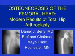
Definition and Prevalence: Bone death is due to either to impaired blood supply or to sever marrow and bone cell damage. • Osteonecrosis is the final common pathway of several diseases entities which result in impaired blood supply to bone tissue causing necrosis and death of the bone. sickle cell disease ,alcohol abuse ,fractures. corticosteroid ,renal disease, diving. • Femoral head is the most vulnerable site for development of AVN characterized by area of death trabecular bone and marrow extending to involve the subchondral plate superior anterolateral aspect of the femoral head ,weight bearing region. • Unknown exact prevalence. • U.S 10.000 - 20.000 new pt./ year. • AVN 10% of THA. • Male / female 8 :1 • Bilateral in younger age group. • Age 40 years. (20 - 50) • History: James Russell – 1794 first to description of bone necrosis, septic. Axhausen -- 1928 AVN occurs in the absence of infection. 1940 AVN occur in arterial occlusion .
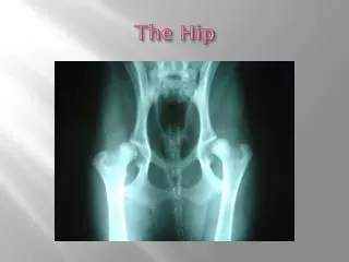
Gross anatomy of femoral head Acetabulum provide coverage 40% of the femoral head (horse shoe ,lunate surface. Fova capitis is a small depression on the medial femoral head is site of attachment of ligament teres. Medial epiphyseal artery from obturator via ligament teres Medial circumflex femoral artery supplying the sup. and inf. Retinocular vessels. Lateral epipheseal vessels enter the femoral head 1cm wide zone b/w the femoral head and the cortical bone of the femoral neck ,supply lateral and central 1/3 F.H Intramedullary vessels from shaft and trochanteric region.
Pathogenesis: • Interruption of the the arterial inflow: post traumatic osteonecrosis is due to interruption of the arterial supply in areas of poor collateral circulation ex: femoral neck fracture ,hip dislocation, slipped epiphysis lead to element of venous tamponade due to intraarticular bleeding and stretching of the capsule. • Venous occlusion: Extra osseous venous occlusion must needs be very extensive to produce vascular stasis and bone ischemia because the communicating diaphseal medullary veins provide such effective collateral drainge(siris &gilfillan 1965 . Venous drainage of femoral head is reduced in Perth's disease (Green&Griffin 1982) • Intravascular capillary occlusion: sickle cell disease clumping of the abnormal red cells may lead to diminished capillary perfusion (Rickles& leary 1974). • Intraosseous capillary tamponade: in alcohol and corticosteroid induced osteonecrosis, the marrow fat cell volume is significantly increase intramedulary pressure is markedly elevated and venous drainage is impaired .( Ficat & arlet 1980)
Sequelae of AVN: • Minimal disease: If the vascular area is small and not adjacent to an articular surface, the patient may be asymptomatic and may heal spontaneously, and the disease may remain undetected or be discovered incidentally during workup for other conditions. • More severe disease: Repair begins at the interface between viable and necrotic bone. Ineffective resorption of dead bone within the necrotic focus is the rule. Reactive and reparative bone is laid down on dead trabeculae resulting in a sclerotic margin of thickened trabeculae within an advancing front of hyperemia, inflammation, bone resorption, and fibrosis. • Mechanical failure: Mechanical failure of trabecular bone at the interface between dead and viable bone may exacerbate AVN. In the subchondral region, such microfractures do not heal because they occur within an area of dead bone. Progression of the microfractures results in a diffuse subchondral fracture, seen radiographically as the crescent sign . Following subchondral fracture and progressive weightbearing, collapse of the articular cartilage occurs .
Conditions associated with osteonecrosis: • Fractures of the femoral neck. • Traumatic hip dislocation. • Slipped capital femoral epiphysis. • Following cervical osteotomy. • Sickle cell anemia. • Alcohol abuse. • Corticosteroid administration. • Rheumatoid arthritis. • Systemic lupus erythematosus. • Chronic pancreatitis. • Radiation. • Renal transplant. • Caisson disease. • Gaucher’s disease. • Pregnancy • Tumors. • Dyschondroplasia. • Coagulative deficiencies. • Cigarette smoking.
Neck Fracture of the femur: Damage to the retinacular vessels supplying the femoral head , and tamponade due to bleeding into confined joint space.( Woodhouse1964, Thompson 1965) Incidence: 16% undisplaced united fracture. 27% displacements fracture (1976 B increase incidence with degree of displacement of the head. (Garden 1961). Traumatic dislocation: Incidence: 30.2% anterior dislocation. 13.4% posterior dislocation. (epstein 1973). 17.6% reduction with two hours. 56.9% prolonged several hours. Slipped capital femoral epiphysis: SCFE is less likely to produce AVN of the epiphysis than a forceful manipulation to reduce the slipped epiphysis (lowe 1961),the capital epiphysis slips posteriorly and therefore dose not damage the posteriorly situated retinacular vessels where as reduction of the slip may do so. Acute cartilage necrosis is thought to occur in 1/3 of patients with SCFE.
Legg-calve- perthes: Most common cause AVN in children. Time onset from 3-10 years. Artery of ligament terse dose not penetrate the epiphysis of the femoral head until age 9-10 years epiphyseal growth plate prevent communication between the blood supply of the epiphysis and the metaphysics. femoral head have high risk to develop AVN even with minor trauma. Steroid administration: possible mechanism: • Occlusion of small vessels occur related to fat emboli from liver. • Increased introsseous pressure result from a steroid related increase in the size of the intramedullary fat cells without an equivalent compensatory loss of the trabecuular and cortical bone. • Fat emboli become hydrolyzed to free fatty acids ,which are toxic to vascular endothelium causing intravascular coagulation. • A direct toxic effect occurs on the osteogenic cells. • Steroid use causes conversion of hematopoietic marrow to fatty marrow leading to AVN.
Clinical presentation: • Pain: • Focal over the groin. • Radiate to buttocks ,anteromedial thigh. • Induced mechanically by standing ,walking. • Throbbing ,deep, often. intermittent. • 40% of patients with night pain associated with morning stiffness. • Click: • A click heard when patient rises from a sitting position • Elicited by external rotation of an abducted hip. • Range of motion: • ROM diminished especially after collapse of femoral head. • ROM limited in flexion ,abduction and internal rotation. • Gait : • walk with limp. • ;
Classification: • Type I : Prenecrotic interstitial edema ,foam cells formation. • Type II : medullary spaces filled with necrotic tissue. • Type III : marrow necrosis , trabecular necrosis up to10%of lacunae empty. Type IV : complete necrosis with dense medullary fibrosis and new bone formation and dead trabeculae. Staging: Numerous staging system. Ficat and Arlet : first 1980 , based primarily upon radiographic findings. Distinguishing between the very early and potentially reversible changes of bone ischemia and the later abnormalities associated with bone death with progressive collapse and articular destruction. Three important factors related to clinical outcome used to stage the disease: clinical presentation. Radiographic assessment of the extent of disease Acetabular cartilage grading.
Ficat classification: Stage o: suspected disease in the contralateral hip when index joint has definitive finding. no clinical symptoms , normal radiographic ,MRI not diagnostic. Stage I: pain is present, ,limited range of motion, internal rotation, abduction. normal radiographic ,MRI diagnostic. Stage II : Clinical signs persist or worsen, Diffuse sclerosis ,cyst on radiographic Crescent line because of subchondral fracture Stage III : Worsening pain ,limping ,limited range of motion in all plans. appearance of sequestrum manifestation on radiographic by a break on the articular margin. Stage IV : same clinical finding on stage III. Early minimum of 2mm circumferentially intact on the A.P and LAT. indicate preservation of joint space. Stage IV: loss of articular cartilage & development of acetabular osteophyte. Radiographic picture of osteoarthritis superimposed on deformed head.
Steinberg staging: Stage 0 : normal ,preclinical and preradiologic. Stage I : pain in the ant. Groin and thigh is common .,limited range ROW. normal radiographic with minimal demineralization. normal bone scan, abnormal MRI. Stage II: clinical signs persist or worsen. Diffuse or localized area of sclerosis and cystic areas. Stage III: Crescent sign, subchondral fracture .no femoral head flatting. Stage IV: Marked collapse and fracture involve the articular surface, segmental flattening of femoral head. Stage V: Resting pain. limited ROM in all plans. developmental of joint space narrowing, acetabular degeneration
Acetabular cartilage grading: Osteonecrosis of the hip is primarily disease of femoral head ,with secondary acetabular involvement resulting from the mechanical damage caused by collapse and subsequent irregularity of the femoral head Dr .Beaule ,Dr.steinberg have demonstrated acetabular involvement with osteonecrosis of femoral head . Acetabular cartilage change in 40 of 41 hips undergo THA. Beaule: Grade I: minimal changes ,localized softening, limited or no break in the surface. Grade II: Area of fissuring and an irregular surface. Grade III: Definite fibrillation with fissuring extending down to subchondral bone. IIIA no osteophytes .IIIB: noncalcified osteophyte in the acetabular fossa. Grade IV: exposure and erosion of subchondral bone. Steinberg: Grade 0 : Normal Grade I : Mild degeneration ,superficial fibrillation ,irregularity of surface. Grade II : Moderate degeneration ,moderate fibrillation ,thinning of cartilage. Grade III :Sever degeneration ,marked fibrillation ,marked thinning of cartilage with areas of complete erosion to bone
Imaging: Plain film radiography: • A.P view. • Frog-leg lateral view ,evaluate superior portion of the femoral head where subchondral abnormalities could be seen. • Normal , many months after symptoms begin. • Earliest sign : mild density change followed by sclerosis and cyst as the disease progress. • Crescent sign (subchondral radiolucency) is evidence of subchondral collapse. • Loss of sphericity and collapse of femoral head. • Joint space narrowing are visible. • Bone scanning: • Technetium 99m bone scanning is useful in patients with: • Negative radiographs • Unilateral symptoms • No risk factors • Increase uptake due to bone turn over at the junction of the dead and the reactive bone, surrounding a cold area.
MRI: • is the most sensitive 91%. • Changes can be seen early in the course of the disease when other studies are negative. • Single density line represent the separation of the normal and ischemic bone. • A second high intensity line (double line sign), hyper vascular granulation tissue. • Replace venography, bone biopsy. • Precaution in interpreting MRI in asymptomatic patients. • Study (23 )steroid treated patient with SLE ,no hip pain and negative radiograph, 8 (35%) have evidence of osteonecrosis of the femoral head on MRI and 6 (26%) have radionuclide bone scanning. Over three years period only (2 )of the( 8) patients with abnormal MRI develop a lesion detectable by hip radiographs. • Surgical treatment on MRI finding with no symptom result in over treatment . • Single photon emission computed tomorgaphy: Initially, SPECT images reflect vascular integrity. Early in the disease, scans may demonstrate an avascular focus in the presence of normal MRI findings unless MRI contrast is used. Collier found a sensitivity of 85% for SPECT. With triple-head high-resolution SPECT, Lee et al found sensitivity to be 97%.
Diagnostic Procedures • Core biopsy and interosseous pressure measurement: • An open biopsy of 10-mm core of bone from the femoral head can be used for diagnosis. • Measurement of interosseous pressure can be obtained before and after biopsy to confirm decompression of intraosseous space. • Venography: • Injection of contrast under image intensification has been used as part of the functional evaluation of bone when measuring intraosseous pressure. • This can be used to confirm presence of the needle within the head and venous congestion.