Download
1 / 41
440 likes | 595 Vues
Physical Examination (continued). Sean Ragain MD. Quick Review. So far, you’ve covered a lot of ground. Let’s look at what you’ve already seen and heard. Inspection, palpation, percussion, auscultation (not necessarily in this order). Exam is modified per patient

E N D
Physical Examination (continued) Sean Ragain MD
Quick Review • So far, you’ve covered a lot of ground. Let’s look at what you’ve already seen and heard. • Inspection, palpation, percussion, auscultation (not necessarily in this order). • Exam is modified per patient • Only time and practice can lead to proficiency!
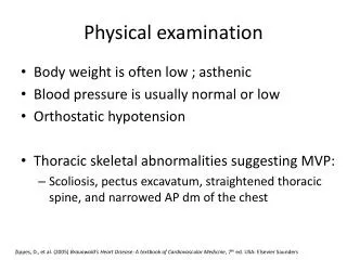
Review • Head exam (HEENT and sometimes N-neck) • Lung topography – imaginary lines, landmarks, fissures, trachea, diaphragm, lung borders • Thoracic Inspection - i.e. barrel chest, pectus excavatum/carinatum, breathing patterns. • Thoracic Palpation – fremitus, crepitus, fractures.
Review • Thoracic Percussion – consolidation? Atelectasis? Increased or decreased resonance?
What’s Next??? • Thoracic Auscultation! Or, auscultation of the lungs. • Mostly done with a stethoscope of course, but you will often get a lot of info just from listening to the person breathe with your “naked” ears. Do your best to achieve quiet in the room so you can hear! • Diaphragm vs. bell of stethoscope. • Mostly, we use the diaphragm, but the bell is particularly good at picking up low frequency sounds, such as certain heart murmurs/valve disorders.
Auscultation • Some sources say to work your way down, but your book says work your way up. Most important: Do side to side comparisons as you move throughout the lung fields.
Stethoscope • What is the difference between the bell and diaphragm? • Diaphragm sits flush against the skin, and therefore filters a lot of low frequency sounds out. • Most lung sounds are high frequency, so we usually employ the diaphragm. • The bell can be an adjunct to the diaphragm. Also, it may be helpful in the emaciated patient. • Don’t press too hard on the bell, this will essentially make it like the diaphragm!
Technique • Quiet Room • As little dress as necessary (and appropriate) • If you must, a thin t-shirt or a hospital gown is ok (but not preferrable) to listen through. • At least one full respiratory cycle at each point of auscultation.
Breath Sounds (table 5-2) • Tracheal breath sounds – Not surprisingly, these should normally be heard over the trachea in the midline of the neck, superior to the sternal notch. They are loud, high pitched, and are equal in length during inspiration and expiration. • Bronchovesicular breath sounds – quieter than tracheal sounds, but otherwise quite similar. Heard around the upper half of the sternum, and between the scapulae on the back. If these sounds are heard coming from large parts of the lung, there may be pathology.
Breath Sounds • Vesicular – soft, muffled sound that is lower in pitch and intensity than tracheal or bronchovesicular breath sounds. Has a longer inspiratory than expiratory component. This is what normal lungs should sound like.
Breath sound intensity • If markedly increased in intensity, they are said to be harsh, the opposite would be described as diminished or even absent.
Adventitious breath sounds • Fortunately, not too many of these to remember, because most diseases present with common adventitious sounds. • Can be classified as continuous or discontinuous. • In general, continuous adventitious sounds, are wheezes, and discontinuous ones are crackles.
Adventitious breath sounds • So we have… • Crackles • Wheezes • Rales • Rhonchi • Stridor • But the terms rales and rhonchi are falling into disuse (to some degree). • Let’s listen to some!
Mechanisms Responsible (intro) • Normal breath sounds are created by turbulent flow in the airways • Bronchial breath sounds heard in the lung periphery may result from consolidation. • Diminished sounds may result from emphysema, collapse, obesity etc. • Crackles are caused by the sudden opening of collapsed airways, or by fluid in the airways. • Wheezes are produced by the vibration of narrowed airways as air passes through (like a reed instrument). • Stridor is often caused by upper airway obstruction.
Examination of the Precordium • The precordium is the surface of the chest wall overlying the heart. • It is examined to assess the condition of the heart. Also, we examine the precordium through the EKG.
Review of Heart Topography • The base of the heart lies directly beneath the middle portion of the sternum • The apex points downward and to the left, extending to the midclavicular line near the 5th ICS.
Inspection and Palpation of the Precordium • Mostly in inspection, you are looking for chest wall deformities, and whether or not you see heaves. What are heaves? • Also, you may try to find the PMI, especially with the initial exam. This may tell you if a person’s heart is enlarged.
Auscultation of heart sounds • The first heart sound (S1) is created by closure of the AV valves. Which ones are those? • The second heart sound (S2) is created by closure of the aortic and pulmonary semilunar valves. • A split S1 or S2 may occur if the opposing valves close at different times. • Significantly split S1 usually indicates a problem (i.e. bundle branch block). • Split S2 can be heard with deep inspiration, but this should return to non-split with exhalation.
Adventitious heart sounds • There are also times when you will hear an S3 or S4. This is often found in heart failure other reasons for the heart to be stiff. • A loud S2 in the “P” area may indicate pulmonary hypertension.
Murmurs • Caused by incompetent or stenotic valves (usually). • Systolic Murmurs are heard when the semilunar valves are stenotic or when the AV valves are incompetent. • Diastolic Murmurs are heard when the AV valves are stenotic or when the semilunar valves are incompetent.
Neurologic Examination • The neurological exam is done by the physician when brain or spinal cord injury is suspected. • RTs need to be familiar with the results of this exam because it has implications for the pulmonary system
Neuro Exam • Review • The CNS is made up of the brain and spinal cord • The brain stem is the most important part of the CNS regarding breathing. It is where breathing is regulated and controlled. • The spinal cord connects the brain to the peripheral body parts for sensory and motor function • The PNS is made up of the cranial nerves and 31 spinal cord nerves
Assessment of the CNS • General level of consciousness • A coma is present when a person cannot be awakened from a sleeplike state. • The Glasgow Coma Scale (GCS) is often used to document the level of neurological impairment. • The patient’s response to pain is also used to assess the CNS. • The patient’s breating pattern often provides clues about the level of brain stem function. Cheyne-Stokes breathing is a common finding with brain stem injury.
Neuro Assessment • PNS • A variety of motor and sensory tests are done. • Reflexes • Strength • Proprioception • Touch, pinprick, vibration. • Cranial Nerves
Abdominal Exam • The proper order of the abdominal exam is more important than the order of the respiratory exam. • Inspection, auscultation, palpation, percussion. • Why?
Abdomen • An enlarged liver is known as hepatomegaly. Right heart failure can cause this. • Severe abdominal distension can impede movement of diaphragm. Such as severe ascites, or bowel obstruction.
Examination of the Extremities • Clubbing • Often seen in patients with chronic cardiopulmonary disease. Cyanotic heart disease, COPD, and lung cancer may also lead to this.
Cyanosis • Peripheral cyanosis is a sign of circulatory disease.
Pedal Edema • May be a sign of chronic lung disease and right heart failure. • But often occurs in healthy older individuals, and may come and go in healthy younger individuals.
Capillary Refill • Will be delayed if cardiac output is poor. • It is tested simply by compressing capillary bed in an extremity, and seeing how long it takes to “pink up”.
Peripheral Skin Temperature • Cool skin is often poorly perfused skin. • THE END