Download
1 / 43
2.14k likes | 8.3k Vues
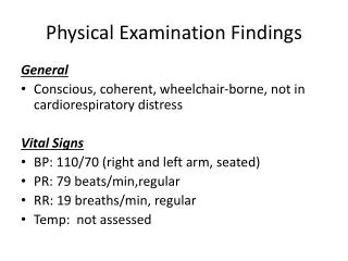
GIT Physical Examination . Hadeel Khadawardi , teaching assistant at Internal Medicine Department, Faculty of Medicine, Umm Al- Qura University. Introduction . Cachectic Jaundiced . General Approach . Vital Signs . Position . Flat On one pillow . GIT Exam . Abdominal Exam .

E N D
GIT Physical Examination HadeelKhadawardi, teaching assistant at Internal Medicine Department, Faculty of Medicine, Umm Al-Qura University
Introduction • Cachectic • Jaundiced General Approach Vital Signs Position • Flat • On one pillow
GIT Exam Abdominal Exam Peripheral Exam • Inspection • Palpation • Percussion (Ascites) • Auscultation • Hand • Arms • Face • Neck: LN • Chest
Hand Nail Palm Dorsum Wrist
Hand • Nail • Clubbing • Peripheral Cyanosis • Leuconychia • Koilonychia What are the causes of clubbing?
Hand • Palm • Pallor • Palmer erythema • Dupuytren contracture
Hand • Dorsum • Muscle Wasting • Tendonousxanthemata
Hand Wrist Flapping tremor(Asterixis) What are the causes of flapping tremor?
Arms • Bruising • Petechiae • Muscle wasting • Scratch marks • Spider nevi How to differentiate between Spider nevi, Venous star & Campbell de Morgan spot?
Face Eyes Salivary Glands Mouth
Face • Eye • Jaundice • Pallor • Xanthelasma • Kayser-Fleischer Ring • ArcusSini • Iritis • PeriorbitalPurpura
Face Parotid gland enlargement What are the causes of unilateral and bilateral parotid gland enlargement?
Face • Mouth • Jaundice • Central cyanosis • Glossitis • Gum hypertophy& pigmentation • Fetor hepaticus • ( methylmerccaptans ) • Mouth ulcers
Lymph Node Examination Neck What is the difference between Troisier’s sign & Trousseau’s sign?
Chest • Gynaecomastia. • Spider Nevi. • Hair Distribution.
Exposure From the nipple to symphysis pubis.
Inspection • (7S) • Symmetrical & movement with respiration. • Scar. • Striae. • Stoma. • Shape of the umbilicus (inverted, flat, exerted). • Shape of the flank (full, straight, empty). • Skin lesions. • (4P) • Prominent veins (caput medusa, SVC obstruction) • Visible Pulsation (aortic aneurysm). • Visible Peristalsis (NL in thin, intestinal obstruction). • Pigmentation (Cullen’s sign, Gery-Turner’s sign) • (1D) • Abdominal Distension (fat, fluid, fetus, flatus, faeces).
Palpation • Before starting palpation, remember: • Relax the abdominal muscles. • If necessary, ask the patient to bend the knee to relax the muscle. • Ask if any particular area is tender and palpate that area last. • Look into patient facial expression while palpating the abdomen.
Palpation Regions of the abdomen
Palpation • Superficial Palpation • Begin with light pressure in each 9 areas. • Start from the Rt. iliac fossa (anti clock wise). • Note the presence of any tenderness or lump.
Palpation • Deep Palpation • Apply more pressure in each 9 areas. • Start from the Rt. iliac fossa (anti clock wise). • Note the presence of any deep tenderness or lump. What is the difference between Guarding and Rigidity?
Palpation • Liver Palpation • Align your hand parallel to the Rt. costal margin, begin in the Rt. Iliac fossa and ask the patient to breath in & out through the mouth. • With each expiration, the hand is moved by 1 or 2 cm closer to the Rt. costal margin. • During inspiration, the hand is kept still waiting for liver edge to strike it.
Palpation • Liver Span • Upper liver border is defined by percussing down at Rt. 2nd IC space in MCL, until dullness is encountered. • Lower liver border is defined by percussing up at Rt. Iliac fossa in MCL, until dullness is encountered. • Measure the distance between the two dull areas. • Normal liver span is 10+/-2.
Palpation • Spleen Palpation • One-hand technique: start from Rt. iliac fossa toward Lt. costal margin and ask the patient to breath in & out through the mouth. With each expiration, the hand is moved by 1 or 2 cm closer to the Lt. costal margin. • Two-hand technique: Lt. hand is placed posterolaterally over Lt. lower ribs and Rt. hand is placed below umbilicus toward Lt. costal margin. • If spleen is not palpable, roll the patient to Rt. Side and palpate again.
Palpation • Spleen percussion • Castell’s Method: percuss on last Lt. IC space & Lt. ant. axillary line. Normally is resonant and dull if splenomegaly. • Traube’s Space: triangle bordered by 6th rib superiorly, Lt. midaxillary line laterally and Lt. costal margin inferiorly. Normally is resonant and dull if splenomegaly. • Nixon’s Method: place the patient on Rt. Lateral decubitus position, percuss at midpoint of Lt costal margin and proceed perpendicularly toward Lt. posterior axillary line. Splenomegaly if there is dullness > 8 cm.
Mild splenomegaly: 1-2 cm below Lt. costal margin. • Moderate splenomegaly: 3-7 cm below Lt. costal margin. • Massive splenomegaly: > 7cm below Lt. costal margin.
Palpation • Kidney palpation • Bimanual palpation (Balloting) • Lt. hand is slide underneath the renal angle. • Flex the fingers at MCP joints to push the content of the abdomen anteriorly. • Place Rt. hand on the top of abdomen at renal angle to palpate for kidney. • Kidney percussion • Kidney is a resonant organ below costal margin.
How to differentiate between splenomegaly & Lt. kidney enlargement ?
Anterior Abdominal Wall Mass • Ask the patient to fold the arms across the upper chest and sit halfway up. If the mass: • Disappear or decrease in size … intra-abdominal mass. • Unchanged … mass is within the abdominal wall.
Percussion • Ascites • Shifting Dullness: • Percuss from the midline out to Lt. flank until dullness is reached. • Mark this point and ask the patient to roll toward you. • Wait for 30 sec. then repeat percuss again. • If the dull area become resonant is indication of ascites. • This maneuver is used to detect mild to moderate ascites.
Percussion • Ascites • Fluid Thrill: • Ask the patient to place the medial edge of his palm firmly on the center of abdomen with fingers directed downward. • Flick the side of abdominal wall and feel the thrill by the other hand on the opposite abdominal wall. • This maneuver is used to detect massive ascites.
Percussion • Ascites • Dipping Maneuver: • To palpate for organomegaly with ascites. • Both hands are placed flat on abdomen and fingers are flexed at MCPs rapidly to displace the underlying fluid.
What is SAAG? What is the DDx of portal HTN & non-portal HTN related ascites?
Auscultation • Bowel Sounds: • Place the diaphragm of stethoscope any where around umbilicus (around iliocecal valve).Percuss from the midline out to Lt. flank until dullness is reached. • Describe it as present or absent. Mark this point and ask the patient to roll toward you. • Absent bowel sounds for 3 min. means paralytic ileus. • Exaggerated bowel sounds mean intestinal obstruction.
Auscultation • Friction rib • Place the diaphragm of stethoscope over liver and spleen. • Hepatic causes: liver tumor, liver abscess, and liver infarction. • Splenic causes: splenic infarction as in SCA, IE.
Auscultation • Venous Hum • Place the bell of stethoscope between xiphisternum & umbilicus. • In portal HTN.
Auscultation • Bruits • ( use bell of stethoscope) • Arterial systolic bruit … over liver … HCC. • Renal bruit … on either side of midline above umbilicus … RAS. • Epigastric bruit … epigastric area … mesenteric artery stenosis.
To complete your GIT exam, exam: • PR • Back • Legs • Genitalia
What are the stigmata of CLD? What are the signs of CLD caused by elevated estrogen level?