Download

1 / 19
230 likes | 272 Vues
Virology. Lab 6. Definitive Properties of Viruses. An infectious, obligate intracellular parasite Viral genome comprised of either DNA or RNA Within an appropriate host cell, the viral genome is replicated and directs the synthesis (utilizing cellular systems) of other viral components

E N D
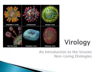
Virology Lab 6
Definitive Properties of Viruses • An infectious, obligate intracellular parasite • Viral genome comprised of either DNA or RNA • Within an appropriate host cell, the viral genome is replicated and directs the synthesis (utilizing cellular systems) of other viral components • Progeny infectious virus particles (virions) are formed by de novo assembly from newly synthesized components within the host cell • A progeny virion assembled during the infectious cycle is the vehicle for transmission of the viral genome to the next host cell or organism, where its disassembly leads to the beginning of the next infectious cycle
Objectives • Introduction to virus culture • Isolation of virus • Quantification of virus
Cell Culture • Cell culture is still the most common method for the propagation of viruses. • Tissues are dissociated into a single-cell suspension by mechanical disruption, followed by treatment with a proteolytic enzyme. • Cells are cultured in a plastic flask in Minimum Essential Media (MEM). As the cells divide, they cover the plastic surface. • Epithelial and fibroblastic cells attach to the surface of the plastic and form a monolayer. • Two commonly used cells in fish virology- EPC - epitholioma papillosum carpio cells- CHSE - chinook salmon embryo
Developing a cell line Live tissue cells to be cultured Culture vessel with appropriate growth media Lift cells into solution with enzyme Cell adhere to vessel and grow to form a monolayer Seed cells into new culture vessels
Isolation of virus from fish • Aseptically remove the internal organs (kidney, liver, spleen, and intestine) and homogenize • Take 0.5g of homogenized tissue and mix with 4.5 ml HBSS to make 10-1 dilution • Make 10-2 dilution of the tissue • Filter the 10-2 dilution with a syringe and Swinney adapter with a 0.45 µm filter. This will trap bacteria in the filter but not virus • Continue making 10-fold dilution series from filtered 10-2 dilution up to 10-5
Virus Dilution 0.1 ml 0.1 ml 0.1 ml 0.1 ml 0.1 ml 1 1/10 1/100 1/1000 1/10000 1/100000 Stock 0.9 ml 0.9 ml 0.9 ml 0.9 ml 0.9 ml 101 10-1 10-2 10-3 10-4 10-5
Cytopathic Effect • Some viruses kill the cells in which they replicate, and infected cells may eventually detach from the cell culture plate. • As more cells are infected, the changes become visible and are called cytopathic effects.
Nuclear shrinking (pyknosis) Proliferation of nuclear membrane Vacuoles in cytoplasm Syncytia (cell fusion) Margination and breaking of chromosomes Rounding up and detachment of cultured cells Inclusion bodies Examples of Cytopathic Effects of Viral Infection
Quantification of CPE • Tissue Culture Infective Dose 50 (TCID50): a measure of virulence of virus • Why Quantify? • Virulence • Immunity • Strain
1 2 3 4 5 6 10-3 10-1 10-4 10-2 10-5 A B 10-6 C 10-7 D Control Multi-well Plates
100 0 Decreasing Dilution TCID50 Procedure • Count wells exhibiting CPE • Ideally you would know all the dilution factors to get infection rates of zero to 100 percent CPE
Calculation of TCID50 • In any biological quantification, the most desirable endpoint is one representing a situation in which half of the inoculated animals or cells show the reaction (death in the case of animals and in CPE case of cells) and the other half do not. • Reed-Muench Method of computing a 50% endpoint of a virus titration • Calculates the proportionate distance between dilutions which infect above and below 50% of the wells
1 2 3 4 5 6 10-1 10-5 A 10-2 10-6 B C 10-3 10-7 D 10-4 Control TCID50 Dilution Infected % Infected10-1 3/3 10010-2 3/3 10010-3 3/3 10010-4 2/3 6610-5 1/3 3310-6 0/3 010-7 0/3 0 Log PD = 66-50 x (Log10) 66-33 Log PD = 0.48Log Dilution above 50 % Infection 10-4.48
Plaque Forming Units • Areas where infected cells are being lysed by virus are seen as plaques, or areas of clearing in the cell monolayer. • When stained with Crystal Violet these areas are easily identified as areas without stain
Plaque forming Units • A single virus infective dose can cause an area of cell destruction • Movement of virus within cell is restricted by an agar overlay • This causes areas of localized destruction • Plaques are enumerated under a microscope to determine the plaque forming units per ml (PFU/ml) • This allows comparison of different viruses in the same unit
Calculation of PFU/mL • Plaques are enumerated • Plaque Counts are averaged over wells • The average is then divided by the dilution times the volume (43+40+38)/3(10-4 x 0.1) = 3,730,000 pfu/ml 43 4 1 040 3 0 038 6 2 0Plaques formed per well
Today’s Lab • Calculate PFU/ml • Calculate TCID50 • Observe infected histology sections • Record values and observations in lab notebook • Observe cell monolayer on inverted scope