Download
1 / 86
960 likes | 1.63k Vues
COPD. COPD. Hallmark symptom - Dyspnea Chronic productive cough Minor hemoptysis pink puffer blue bloater. COPD- pulmonary hyperinflation- the diaphragms are at the level of the eleventh posterior ribs and appear flat. COPD - Physical Findings. Tachypnea Accessory respiratory muscle use

E N D
COPD • Hallmark symptom - Dyspnea • Chronic productive cough • Minor hemoptysis • pink puffer • blue bloater
COPD- pulmonary hyperinflation- the diaphragms are at the level of the eleventh posterior ribs and appear flat.
COPD - Physical Findings • Tachypnea • Accessory respiratory muscle use • Pursed lip exhalation • Weight loss due to poor dietary intake and excessive caloric expenditure for work of breathing
Dominant Clinical Forms of COPD • Pulmonary emphysema • Chronic bronchitis • Most patients exhibit a mixture of symptoms and signs
COPD - Advanced Dx • secondary polycythemia • cyanosis • tremor • somnolence and confusion due to hypercarbia • Secondary pulmonary HTN w or w/o cor pulmonale
COPD Treatment Strategy • Elimination of extrinsic irritants • bronchodilator & glucocorticoid therapy • Antibiotics • Mobilization of secretions • “respiratory vaccines” • Oxygen therapy - if oxygen saturation <90% at rest on room air
A-a gradient A-a gradient = predicted pO2 – observed PO2 PAO2 = (FIO2 X 713) – (PaCO2/0.8) at sealevel PAO2 = 150-(PaCO2/0.8) at sealevel on room air Normal range 10-15mm > 30 years of age Normal range 8mm < 30 years of age Increased A-aDO2=diffusion defect Right to left shunt V/Q mismatch
Examples • A doubel overdose brings two 30 yr old patients to the ED. Both have ingested substantial amounts of barbiturates and diazepam. Blood gases drawn on room air revealed these values: • patient 1- pH =7.18, PCO2 = 70mmHg, PO2=50mmHg, HCO3=24mEq/L; • patient2- pH =7.31, PCO2=50mmHg, PO2=50mmHg, HCO3=25mEq/L
Comment • The A-a gradient calculation for patient 1 is as follows: • A-a DO2 = PAO2 – PaO2 • PAO2 = 150 – (1.25x PCO2) • PAO2 = 150 – (1.25x 70) • PAO2 = 62 • A-a =62 – 50 • A-a = 12
Comment • The calculation reveals a normal gradient, indicating that the etiology for hypoxemia and hypoventilation is extrinsic to the lung itself.
Comment • The A-a gradient calculation for patient 2 is as follows: • PAO2 = 150 – (1.25 x PCO2) • PAO2 = 150 – (1.25 x 50) • PAO2 = 150 – 63 • PAO2 = 87 • Therefore, A-a = 87 – 50 =37 (an abnormally increased gradient)
Comment • We can be reasonably confident that patient 1 suffered hypoventilation due to the effect of the ingested drugs on the brain stem. • Temporary mechanical ventilation restored this patient’s gas exchange.
Comment • Patient 2, on the other hand, had an increased A-a gradient, indicating a lung problem in addition to any central cause for hypoventilation. • The chest x-ray film revealed that this patient’s overdose was complicated by aspiration pneumonitis and that the patient required treatment with antibiotics in addition to mechanical ventilation.
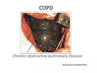
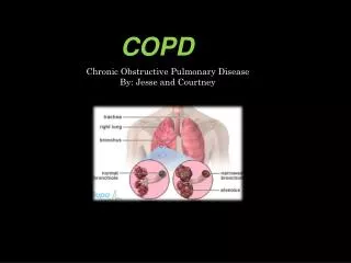
(Chronic Obstructive Pulmonary Disease) COPD • Chronic Bronchitis • Emphysema
Definition • A disease state characterized by airflow limitation that is not fully reversible • Conditions include: • Emphysema: (anatomically defined condition characterized by destruction and enlargement of the lung alveoli) • Chronic bronchitis: clinically defined condition with chronic cough and phlegm • Small-airways disease: condition in which small bronchioles are narrowed
Epidemiology • Fourth leading cause of death in the U.S. • Affects > 16 million persons in the U.S. • Global Initiative for Chronic Obstructive Lung Disease (GOLD) estimates suggest that chronic obstructive lung disease (COLD) will increase from the sixth to the third most common cause of death worldwide by 2020.
Epidemiology • >70% of COLD-related health care expenditures go to emergency department visits and hospital care (>$10 billion annually in the U.S.).
Epidemiology Sex • Higher prevalence in men, probably secondary to smoking • Prevalence of COLD among women is increasing as the gender gap in smoking rates has diminished.
Epidemiology Age • Higher prevalence with increasing age • Dose–response relationship between cigarette smoking intensity and decreased pulmonary function
Risk Factors • Cigarette smoking is a major risk factor. • Cigar and pipe smoking • Passive (secondhand) smoking Associated with reductions in pulmonary function Its status as a risk factor for COLD remains uncertain
Occupational exposures to dust and fumes (e.g., cadmium) • Likely risk factors • The magnitude of these effects appears substantially less important than the effect of cigarette smoking. • Ambient air pollution • The relationship of air pollution to COLD remains unproven.
Genetic factors • α1 antitrypsin (α1AT) deficiency • Common M allele: normal levels • S allele: slightly reduced levels • Z allele: markedly reduced levels • Null allele: absence of α1AT (rare) • Lowest levels of α1AT are associated with incidence of COLD; α1AT deficiency interacts with cigarette smoking to increase risk.
Distributions of forced expiratory volume in 1 s (FEV1)values in a generalpopulation sample, stratified by pack-years of smoking
EtiologyCOLD • Causal relationship between cigarette smoking and development of COLD has been proven: however, the response varies considerably among individuals.
COLD exacerbation • Bacterial infections Streptococcus pneumoniae Haemophilus influenzae Moraxella catarrhalis Mycoplasma pneumoniae or Chlamydia pneumoniae (5–10% of exacerbations) • Viral infections (one-third) • No specific precipitant identified (20–35%)
Symptoms & Signs • 3 most common: • Cough • Sputum production • Exertional dyspnea, frequently of long duration
signs and symptoms • Dyspnea at rest • Prolonged expiratory phase and/or expiratory wheezing on lung examination • Decreased breath sounds • Barrel chest • Large lung volumes and poor diaphragmatic excursion, as assessed by percussion • Use of accessory muscles of respiration • Pursed lip breathing (predominantly emphysema) • Characteristic "tripod" sitting position to facilitate the actions of the sternocleidomastoid, scalene, and intercostal muscles • Cyanosis, visible in lips and nail beds
Systemic wasting Significant weight loss Bitemporal wasting Diffuse loss of subcutaneous adipose tissue Paradoxical respiration Inward movement of the rib cage with inspiration (Hoover's sign) in some patients "Pink puffers" are patients with predominant emphysema—no cyanosis or edema, with decreased breath sounds. "Blue bloaters" are patients with predominant bronchitis—cyanosis and edema. Most patients have elements of each.
Advanced disease: signs of cor pulmonale Elevated jugular venous distention Right ventricular heave Third heart sound Hepatic congestion Ascites Peripheral edema
Differential Diagnosis • Congestive heart failure • Asthma • Bronchiectasis • Obliterative bronchiolitis • Pneumonia • Tuberculosis • Atelectasis • Pneumothorax • Pulmonary embolism
Considerations • COLD is present only if chronic airflow obstruction occurs. Chronic bronchitis without chronic airflow obstruction is not COLD. • Asthma Reduced forced expiratory volume in 1 second (FEV1) in COLD seldom shows large responses (>30%) to inhaled bronchodilators, although improvements up to 15% are common. Asthma patients can also develop chronic (not fully reversible) airflow obstruction.
Considerations • Problems other than COLD should be suspected when hypoxemia is difficult to correct with modest levels of supplemental oxygen. • Lung cancer Clubbing of the digits is not a sign of COLD.In patients with COLD, development of lung cancer is the most likely explanation for newly developed clubbing.
Chronic Bronchitis • Chronic lower airway inflammation • Increased bronchial mucus production • Productive cough • Urban male smokers > 30 years old
Chronic Bronchitis • Mucus, swelling interfere with ventilation • Increased CO2, decreased 02 • Cyanosis occurs early in disease • Lung disease overworks right ventricle • Right heart failure occurs • RHF produces peripheral edema Blue Bloater
Emphysema • Loss of elasticity in small airways • Destruction of alveolar walls • Urban male smokers > 40-50 years old
Emphysema • Lungs lose elastic recoil • Retain CO2, maintain near normal O2 • Cyanosis occurs late in disease • Barrel chest (increased AP diameter) • Thin, wasted • Prolonged exhalation through pursed lips Pink Puffer
COPD Management • Oxygen • Monitor carefully • Some COPD patients may experience respiratory depression on high concentration oxygen • Assist ventilations as needed
Diagnostic Approach Initial assessment • History and physical examination (Signs & Symptoms) • Pulmonary function testing to assess airflow obstruction • Radiographic studies
Assessment of exacerbation • History Fever Change in quantity and character of sputum ill contacts Associated symptoms Frequency and severity of prior exacerbations
Assessment of exacerbation • Physical examination Tachycardia Tachypnea Chest examination Focal findings Air movement Symmetry Presence or absence of wheezing Paradoxical movement of abdominal wall Use of accessory muscles Perioral or peripheral cyanosis Ability to speak in complete sentences Mental status
Radiographic studies Chest radiography focal findings (pneumonia, atelectasis) • Arterial blood gases Hypoxemia Hypercapnia • Hospitalization recommended for: Respiratory acidosis and hypercarbia Significant hypoxemia Severe underlying disease Living situation not conducive to careful observation and delivery of prescribed treatment
ABG and oximetry • Although not sensitive, they may demonstrate resting or exertional hypoxemia. • Blood gases provide additional information about alveolar ventilation and acid–base status by measuring arterial PCO 2 and pH. • Change in pH with PCO 2 is 0.08 units/10 mmHg acutely and 0.03 units/10 mmHg in the chronic state.
Laboratory Tests • Elevated hematocrit suggests chronic hypoxemia. • Serum level of α1AT should be measured in some patients. o Presenting at ≤ 50 years of age o Strong family history o Predominant basilar disease o Minimal smoking history o Definitive diagnosis of α1AT deficiency requires PI type determination. Typically performed by isoelectric focusing of serum, which reflects the genotype at the PI locus for the common alleles and many of the rare PI alleles Molecular genotyping can be performed for the common PI alleles (M, S, and Z). • Sputum gram stain and culture (for COLD exacerbation)
Imaging • Chest radiography • Emphysema: obvious bullae, paucity of parenchymal markings, or hyperlucency • Hyperinflation: increased lung volumes, flattening of diaphragm • Does not indicate chronicity of changes • Chest CT • Definitive test for establishing the diagnosis of emphysema, but not necessary to make the diagnosis
Diagnostic Procedures Pulmonary function tests/spirometry • Chronically reduced ratio of FEV1 to forced vital capacity (FVC) • In contrast to asthma, the reduced FEV1 in COLD seldom shows large responses (>30%) to inhaled bronchodilators, although improvements up to 15% are common. • Reduction in forced expiratory flow rates • Increases in residual volume • Increases in ratio of residual volume to total lung capacity • Increased total lung capacity (late in the disease) • Diffusion capacity may be decreased in patients with emphysema. Electrocardiography • may detect signs of ventricular hypertroph
Classification • GOLD stage • Classification based on pathologic type