Download
1 / 15
200 likes | 1.03k Vues
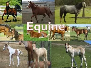
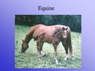
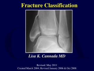
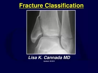
Equine P3 Fracture Classification. James Montgomery, DVM June 1, 2009. Deuce Acc # 109775. 7 year old Female Quarter horse Hx: Possible P3 fracture, right hind. Deuce Acc # 109775. Parasagittal (Type II) fracture of distal phalanx. Deuce Acc # 109775. Classification system.

E N D
Equine P3 Fracture Classification James Montgomery, DVM June 1, 2009
Deuce Acc # 109775 • 7 year old • Female • Quarter horse • Hx: Possible P3 fracture, right hind
Deuce Acc # 109775 • Parasagittal (Type II) fracture of distal phalanx
Classification system • Type I- Non-articular of Palmar or plantar process • Type II- Articular fxs from distal interphalangeal joint to solar margin • Type III- Articular midsagittal fx – divides into equal parts • Type IV- Articular fracture - Extensor process • Type V- Articular comminuted body fx • Type VI- Solar margin only • Type VII – Palmar process in the foal
P3 Fractures • Trauma most common cause • Type VI fractures more common than the other 5 types combined • Make sure 65◦ DP radiographs are not overexposed may miss Type VI fractures
Fracture healing • Difficult to assess progression radiographically – minimal amount of callus formation • Fractures of extensor process produce the greatest amount of new bone • Treatment by corrective shoeing and stall rest healing in 3 to 19 months • Young horses and nonarticular fractures show most rapid and complete progression to bone union
Prognosis • Return to athletic activity is good for type I • Guarded for types II and IV • Solar margin (type VI) fractures have a good prognosis if not associated with laminitis or severe pedal osteitis
Differentiating fracture from artifact • Both and acute Type II or III fracture and packing artifact can appear as a linear lucent finding • 3 factors to help differentiate • Clinical signs – fracture usually seriously lame • Fracture will extend to and end at the margins of P3 – packing artifact may extend beyond the margins of P3 • Linear lucent packing artifacts extend obliquely from the heel (abaxially) toward the toe (axially) • Type II fractures run obliquely from the solar margin of the quarter (abaxially) towards the articulation (axially) • Type III fractures run sagitally from the toe to the articulation
Palmar Process Fractures in the Foal • Ossicles of the medial and/or lateral palmar and plantar processes of P3 are seen in foals from a few weeks to a year of age • These are Type VII fractures rather than secondary centers of ossification or developmental orthopedic disease (DOD) • May be seen in club-footed foals or in foals with or without clinical signs of lameness
Palmar Process Fractures in the Foal • Two radiographic patterns associated with this type of fracture are: • Triangular bony fragment at the palmar aspect of the distal angle of the palmar process • Oblong fragment separated from P3 by a lucent line extending 1-3 from the incisure of the palmar process to the solar margin • Healing radiographically complete in an average of 8 weeks with return to being sound
References • Pack L. Equine Imaging. VCA 341 lecture, Atlantic Veterinary College, University of Prince Edward Island. • Riedesel EA. The Phalanges. In Thrall DE, ed. Textbook of Veterinary Diagnostic Radiology, 5th ed (St. Louis, MO: Saunders Elsevier, 2007) pp. 432-3.