Download
1 / 28
280 likes | 497 Vues
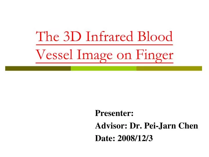
The 3D Infrared Blood Vessel Image on Finger . Presenter: Advisor: Dr. Pei-Jarn Chen Date: 2008/12/3. Outline. Introduction Papers Review Motivation and Purpose Materials and Methods Preliminary Results Future Works References. Introduction. Diabetes in Taiwan

E N D
The 3D Infrared Blood Vessel Image on Finger Presenter: Advisor: Dr. Pei-Jarn Chen Date: 2008/12/3
Outline • Introduction • Papers Review • Motivation and Purpose • Materials andMethods • PreliminaryResults • Future Works • References
Introduction • Diabetes in Taiwan • Among the population over 65 years old • 20% of they are diabetic • Ten major causes of death in 2007 • http://www.doh.gov.tw/ Diabetes 糖尿病
Introduction • What is diabetes? • Diabetes is a disease in which the body does not produce or properly use insulin. • Type I • Type II • Type III • Other • Complication of Diabetes • Pathological change • Nerve • Eyes • Heart vessel • Kidney • Infection of the foot Eyes Pathological change Infection of the foot pathological change Diabetes 糖尿病
Introduction • Inspection • Computed Tomography( CT ) • Magnetic Resonance Imaging (MRI) • Digital Subtraction Angiography( DSA ) 電腦斷層掃描 磁振造影 數位減法血管攝影 CT MRI DSA
Paper Reviews(1) • Comparison of Major Biometrics Methods ◎: good, ○: normal, × : insufficient Junichi Hashimoto,” Finger Vein Authentication Technology and its Future”, IEEE Trans. Med. Imaging ,2006.
Paper Reviews(1) • Finger Vein Imaging Method Light Reflection Method Light Transmission Method Junichi Hashimoto,” Finger Vein Authentication Technology and its Future”, IEEE Trans. Med. Imaging ,2006.
Paper Reviews(2) • Optical window used for detecting the vein pattern oxidized hemoglobin [HbO] 含氧血紅素 deoxidized hemoglobin [Hb] 去氧血紅素 Septimiu Crisan, Joan Gavril Tarnovan, and Titus Eduard CriUan,“A Low Cost Vein Detection System Using Near Infrared Radiation”, IEEE Trans. Biomed. Eng ,2007.
Paper Reviews(2) • Why do we select the hand? • It is usually an uncovered part of the body and it has sufficient mobility. • It has a reduced amount of hair especially in the front of the hand. • The veins are closer to the surface than in other parts of the body and therefore easier to scan. • The vein pattern is intricate enough to allow sufficient criteria for positively detecting various subjects even identical twins. Septimiu Crisan, Joan Gavril Tarnovan, and Titus Eduard CriUan,“A Low Cost Vein Detection System Using Near Infrared Radiation”, IEEE Trans. Biomed. Eng ,2007. 9
Motivation and Purpose • To design a low-cost and an easy-to-use 3D image system on the finger extremity and personal databases.
Materials andMethods • System Flow Chart
Materials andMethods 003 002 001 001:攝像系統包括鏡頭與濾鏡。 002:為觀察者手指放置基座。 003:為一組平面之紅外線LED光源。 004:紅外線LED燈亮度調節器 005:為2組齒輪。 006:為ㄧ步進馬達。 007:步進馬達的控制電路 LED燈 亮度調節器 步進馬達 控制電路 004 005 006 007
Materials andMethods • Camera Module • Camera- UI-1220 • 752*480 • bmp • Lens- IDS-10 • Filter Lens- B+W infrared Filter 093 • Light Source- IR LED • NoteBook • 1.66 GHz • 512 MB DDR RAM • Windows XP • MATLAB • LABView
Materials andMethods • Stepper Motor (KH42HM2 B021) 六線式步進馬達, 精度1.8 DEG, 兩相激磁 Stepper Motor 步進馬達
Line integral projection P(p,q) of the two-dimensional Radon transform. y q p f(x,y) q x p P(p,q) q Materials andMethods • Radon Transform 上式的就是所謂f(x,y)的Radon Transform 15
Materials andMethods • Fourier Slice Theorem 將上面固定角度所得到的 ,經過傅立業轉換,定義成 代入 得到 將 轉換回(x,y)座標系統得到 上式說明 可看成為f(x,y)在 的二維傅立業轉換 即 16 Fourier Slice Theorem 傅立業切片定理
Materials andMethods • Filtered Back-Projection • (a)藍姆-拉克 (Lam-Sak) • (b)謝卜-羅庚 (Shepp-Logan) • (c)低通餘弦 (low pass cosine) • (d)通用漢米 (hamming) 17 Filtered Back-Projection 濾波反投影
Materials andMethods • Filtered Back-Projection 因此經過濾波後之 其數學表示式如下: 而物體函數f(x,y)則為 綜合上述原理,整個平形光束反投影演算法之步驟整理如下: (1)定義投影角度之個數 K 與每次投影之角度,其中0< θ < 180° (2)測量投影, (3)尋找每一投影函數(即Radon轉換), (4)乘以加權因子 (5)加總布満影像平面濾波投影後之反傅立業轉換(反投影處理) 18
Materials andMethods 3D影像疊成 利用不同角度之平行 光束的投影的Radon 轉後,再經反傅立業 轉換重建得到每一切 面之斷層影像後,接 著我們只要把每一切 面之邊緣對齊或利用 連續曲線密合(fitting) ,就可以得到我們想 要的3D手指血管影像 19
Preliminary Results Finger Vein Finger vein acquisition module Finger Vein
Preliminary Results • User Interface
Preliminary Results • Different Anglesof Images 0° 45° 90° 135° 180°
Future Works • To distinguish arterial from vein on finger using developed system. • To reconstruction the 3D image using developed system.
References • 行政院衛生署(Department of Health,Executive Yuan, Taiwan, R.O.C)http://www.doh.gov.tw/ • 鍾豐橋,人體手足末端血管紅外線影像擷取與量化分析系統之建立,南台科技大學電機工程研究所碩士學位論文,2007。 • Junichi Hashimoto,” Finger Vein Authentication Technology and its Future”, IEEE Trans. Med. Imaging,2006. • Septimiu Crisan, Joan Gavril Tarnovan, and Titus Eduard CriUan,“A Low Cost Vein Detection System Using Near Infrared Radiation”, IEEE Trans. Biomed. Eng,2007. • Moulay Akhloufi, Abdelhakim Bendada,“Hand and wrist physiological features extraction for near infrared biometrics”, 2008.
Thank you for your attention.
Pre-processing Original Image Image Enhancement Median Filter Average Filter Binary Morphology Remove Small Regions
所謂斷層掃描( tomography) , 是以大量經過物體截面 • 的衰減訊號來產生斷面影像。早在1917 年Radon 發表可以 • 從投影 ( projections ) 數據重建原函數的理論( Radon, • 1917),即已奠定斷層掃描成像的數學基礎( Radon, 1917; • Deans, 1983;Kak and Slaney, 1988)。傅氏切片定理( Fourier • slice theorem)( Bracewell, 1956)告訴我們:物件函數的二 • 維傅立葉轉換空間被每一個測量投影所佈滿,唯有當傅立葉 • 空間完全佈滿時,才有希望找到唯一的物件函數。斷層掃描 • 成像技術如前所述是利用數學上的 Radon 轉換和傅氏光學 • 的理論發展出來的,結合影像重建理論和電腦高速的運算能 • 力使得產生高解析度的斷面影像更為可行。