Download

1 / 44
550 likes | 886 Vues
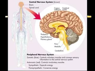
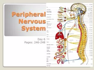
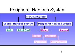
Welcome To PERIPHERAL NERVOUS SYSTEM. Functional Organization of the PNS. Nervous structures outside the brain and spinal cord - sensory and motor connections to the outside world - nerves thread throughout the body to allow the CNS to receive information and take action

E N D
Welcome To PERIPHERAL NERVOUS SYSTEM
Nervous structures outside the brain and spinal cord - sensory and motor connections to the outside world - nerves thread throughout the body to allow the CNS to receive information and take action • Functional components of the PNS - sensory inputs and motor outputs - categorized as somatic or visceral - also classified as general or special
Functional Components of the PNS • Basic structural components: 1. Sensory receptors – pick up stimuli from inside and outside the body, then initiate impulses in sensory axons 2. Motor endings – the axon terminals of motor neurons that innervate the effectors 3. Nerves – bundles of peripheral axons and Ganglia - clusters of peripheral neuronal cell bodies - most are mixed nerves, contain both sensory and motor axons - some cranial nerves are purely sensory or purely motor in function
3 main types of nerve cells sensory neurone relay neurone/interneuron motor neurone
Sensory neurons Carries impulses from receptors e.g pain receptors in skin to the CNS( brain or spinal cord)
Relay neuron/ Interneurons Carries impulses from sensory nerves to motor nerves.
Motor neuron Carries impulses from CNS to effector e.g. muscle to bring about movement or gland to bring about secretion of hormone e.g ADH
Spinal nerves Dorsal roots – sensory fibers arising from cell bodies in dorsal root ganglia Ventral roots – motor fibers arising from anterior gray column of spinal cord
Autonomic Nervous System • The autonomic nervous system is the subdivision of the peripheral nervous system that regulates body activities that are generally not under conscious control • General visceral motor part of the PNS • Has 2 divisions (with opposite effects): - Parasympathetic: ‘housekeeping’ activities (rest and digest) - Sympathetic: extreme situations (fight or flight)
ANS is the subdivision of the peripheral nervous system that regulates body activities that are generally not under conscious control • Visceral motor innervates non-skeletal (non-somatic) muscles • Composed of a special group of neurons serving: • Cardiac muscle (the heart) • Smooth muscle (walls of viscera and blood vessels) • Internal organs • Skin
Basic anatomical difference between the motor pathways of the voluntary somatic nervous system (to skeletal muscles) and those of the autonomic nervous system • Somatic division: • Cell bodies of motor neurons reside in CNS (brain or spinal cord) • Their axons (sheathed in spinal nerves) extend all the way to their skeletal muscles • Autonomic system: chains of two motor neurons • 1st = preganglionic neuron (in brain or cord) • 2nd = gangionic neuron (cell body in ganglion outside CNS) • Slower because lightly or unmyelinated (see next diagram)
Axon of 1st (preganglionic) neuron leaves CNS to synapse with the 2nd (ganglionic) neuron • Axon of 2nd (ganglionic) neuron extends to the organ it serves Diagram contrasts somatic (lower) and autonomic: autonomic this dorsal root ganglion is sensory somatic Note: the autonomic ganglion is motor
Divisions of the autonomic nervous system (visceral motor part of it) • Parasympathetic division • Sympathetic division
Divisions of the autonomic nervous system • Parasympathetic division • Sympathetic division Serve most of the same organs but cause opposing or antagonistic effects Parasysmpathetic: routine maintenance “rest &digest” Sympathetic: mobilization & increased metabolism “fight, flight or fright” or “fight, flight or freeze”
Where they come from Sympathetic/ thoracolumbar: Thoracic and Lumbar Vertebrae Parasympathetic/ Craniosacral: Cervical (neck area) Caudal (tailbone) Brain (cranial nerves)
Parasympathetic nervous system“rest & digest” • Also called the craniosacral system because all its preganglionic neurons are in the brain stem or sacral levels of the spinal cord • Cranial nerves III,VII, IX and X • In lateral horn of gray matter from S2-S4 • Only innervate internal organs (not skin) • Acetylcholine is neurotransmitter at end organ as well as at preganglionic synapse: “cholinergic”
Parasympathetic continued • Cranial outflow • III - pupils constrict • VII - tears, nasal mucus, saliva • IX – parotid salivary gland • X (Vagus n) – visceral organs of thorax & abdomen: • Stimulates digestive glands • Increases motility of smooth muscle of digestive tract • Decreases heart rate • Causes bronchial constriction • Sacral outflow (S2-4): form pelvic splanchnic nerves • Supply 2nd half of large intestine • Supply all the pelvic (genitourinary) organs
Parasympathetic (only look at this if it helps you)
Sympathetic nervous system“fight, flight or fright” • Also called thoracolumbar system: all its neurons are in lateral horn of gray matter from T1-L2 • Lead to every part of the body (unlike parasymp.) • Easy to remember that when nervous, you sweat; when afraid, hair stands on end; when excited blood pressure rises (vasoconstriction): these sympathetic only • Also causes: dry mouth, pupils to dilate, increased heart & respiratory rates to increase O2 to skeletal muscles, and liver to release glucose • Norepinephrine (aka noradrenaline) is neurotransmitter released by most postganglionic fibers (acetylcholine in preganglionic): “adrenergic”
Sympathetic nervous system continued • Regardless of target, all begin same • Preganglionic axons exit spinal cord through ventral root and enter spinal nerve • Exit spinal nerve via communicating ramus • Enter sympathetic trunk/chain where postganglionic neurons are • Has three options…
Options of preganglionic axons in sympathetic trunk (see next slides for drawing examples) • Synapse on postganglionic neuron in chain ganglion then return to spinal nerve and follow its branch to the skin • Ascend or descend within sympathetic trunk, synapse with a posganglionic neuron within a chain ganglion, and return to spinal nerve at that level and follow branches to skin • Enter sympathetic chain, pass through without synapsing, form a splanchnic nerve that passes toward thoracic or abdominal organs • These synapse in prevertebral ganglion in front of aorta • Postganglionic axons follow arteries to organs
Sensory Receptors of the PNS • Also classified according to: a) Location – based on body location or location of stimuli to which they respond b) Type of stimulus detected – kinds of stimuli that most readily activate them c) Structure – divided into 2 broad categories free or encapsulated nerve endings
Classification by Location • Exteroceptors – sensitive to stimuli arising from outside the body - located at or near body surfaces - include receptors for touch, pressure, pain, temperature, and most receptors of the special sense organs • Interoceptors (visceroceptors) – receive stimuli from internal viscera (digestive tube, bladder, lungs) - monitor a variety of stimuli such as changes in chemical concentration, taste stimuli, stretching of tissues, and temperature - activation causes visceral pain, nausea, hunger, or satiety
Classification by Location • Proprioceptors – monitors degree of stretch and sends input on body movements to the CNS - located in musculoskeletal organs such as skeletal muscles, tendons, joints, and ligaments
Classification by Stimulus Detected • Mechanoreceptors – respond to mechanical forces - such as touch, pressure, stretch, vibrations, and itch • Thermoreceptors – respond to temperature changes • Chemoreceptors – respond to chemicals in solution (molecules tasted or smelled) and to change in blood chemistry • Photoreceptors in the eye – respond to light • Nociceptors – respond to harmful stimuli that result in pain (noci = harm)
Classification by Structure • General sensory receptors – widely distributed • Nerve endings of sensory neurons moniter: - Touch - Pressure - Vibration - Stretch - Pain - Temperture - Proprioception
Classification by Structure • General sensory receptors are divided into 2 groups - Free nerve endings - Encapsulated nerve endings Note: there is no perfect ‘one receptor – one function’ - one receptor can respond to several kinds of stimuli and different receptor types can respond to similar stimuli
Free Nerve Endings • Abundant in epithelia and underlying CT • Respond to pain and temperature • Monitor affective senses – those to which people have an emotional response (pain) • 2 specialized types of free nerve endings: - Merkel discs: lie in the epidermis - Hair follicle receptors: wrap around hair follicles
Free Nerve Endings • Merkel discs – a disc-shaped epithelial cell innervated by a sensory nerve ending - slowly adapting receptors for light touch (respond and send out action potentials even after continual stimulation) • Hair follicle receptors – receptors for light touch - monitor the bending of hairs - rapidly adapting (sensation disappears quickly even if the stimulus is maintained) • Itch receptor – in the dermis (newly discovered)