Download
1 / 39
400 likes | 665 Vues
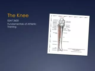
The Knee. From the Sports Medicine Perspective. Bony Anatomy. Femur Patella Tibia Fibula. Bony Anatomy. Femur: Longest Bone in Body Tibia: WB bone of lower extremity Fibula: Site of Muscle Attachment Patella: Sesamoid Bone A bone that develops within a tendon. Knee Skeletal.

E N D
Bony Anatomy Femur Patella Tibia Fibula
Bony Anatomy • Femur: Longest Bone in Body • Tibia: WB bone of lower extremity • Fibula: Site of Muscle Attachment • Patella: Sesamoid Bone • A bone that develops within a tendon
Knee Skeletal Lateral Condyle Head of Fibula Femoral Groove Gerdy’s Tubercle TibialTuberosity Pes Anserine
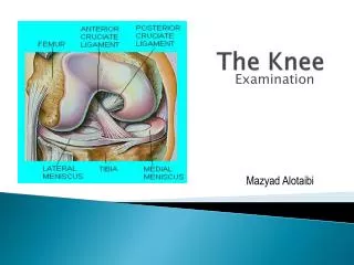
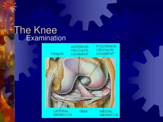
Menisci Medial Meniscus Lateral Meniscus PCL ACL
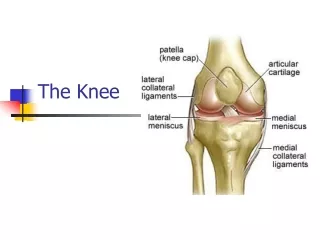
MCL • Thick Band of Tissue • Tibia Femur • Resists Valgus Force
Valgus • Outside to Inside Force • MCL resists this force • Occurs in FRONTAL PLANE
LCL • Narrow cord like band of tissue • Fibula Femur • Resists Varus Forces
Varus • Inside to Outside Force • LCL resists this force • FRONTAL PLANE
Anterior Cruciate Ligament ACL • Composed of 3 bands • Prevents anterior translation of tibia • Stabilizes against excessive rotation • Stabilizing Ligament
Posterior Cruciate Ligament PCL • Stabilizes the posterior aspect of knee • Prevents hyperextension
Quadriceps • Anterior Thigh Musculature • Four Muscles: • Rectus Femoris • Vastus Lateralis • Vastus Medialis • Vastus Intermedius • Extend the Knee
Rectus Femoris • 2 Joint Muscle • Crosses hip and knee • Flexes Hip • Extend the knee • Converges with rest of quadriceps muscles at tibial tubercle
Hamstrings • Three Muscles • Semimembranosus • Semitendinosus • Biceps Femoris • Common Origin the ischial tuberosity • Flex the Knee
MCL Sprains • Valgus Force • Tensile Mechanism MCL • Flexed knee more vulnerable (open pack position = less stable)
MCL Injuries • Direct trauma in frontal plane injures MCL • Combination of rotation can result in ACL and meniscus tears
MCL/LCL Injuries • GRADE I: • No instability • Mild Effusion • ROM full • Mild tenderness w/ palpation
GRADE II: Laxity w/ valgus or varus stress (more with 30 degrees of flexion) Decrease in ROM Increase medial (MCL) or lateral (LCL) pain GRADE III: Complete ligament rupture Complete loss of stability Immediate pain that transitions into dull ache MCL/LCL Injuries
Treatment • Based on severity of injury • RICE • Modify activity • Crutches • Exercises in sagittal plane • Progress to functional exercise