Download
1 / 28
290 likes | 580 Vues
Functional Magnetic Resonance Imaging. Carol A. Seger Psychology Molecular, Cellular, and Integrative Neuroscience Michael Thaut Music, Theater, and Dance and MCIN. Outline. Overview of fMRI Our lab’s research questions Open imaging issues in fMRI

E N D
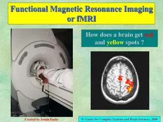
Functional Magnetic Resonance Imaging Carol A. Seger Psychology Molecular, Cellular, and Integrative Neuroscience Michael Thaut Music, Theater, and Dance and MCIN
Outline • Overview of fMRI • Our lab’s research questions • Open imaging issues in fMRI • Spatial normalization and interindividual comparisons • Functional connectivity analyses
fMRI: what are we measuring? BOLD imaging • Blood oxygenation level dependent contrast. • Ratio of deoxyhemoglobin to oxyhemoglobin • Essentially reflects blood flow (hemodynamic response) • Hemodynamic response characteristics • Tightly coupled to neural activity. • Slow • Additive • Inherently comparative method
Design Image acquisition Anatomical images Functional images Across multiple tasks Preprocessing Slice timing correction Temporal smoothing Motion correction Spatial smoothing Normalization to template brain Statistical analyses Deconvolution of BOLD signal Voxel wise statistical analyses comparing BOLD signal to task Correction for multiple comparisons Functional connectivity analyses Data visualization False color overlay onto anatomical images Cortex inflation Steps in fMRI
Introduction to my research questions • “The roles of corticostriatal loops in human learning and cognition” • Corticostriatal loops and the basal ganglia • Human stimulus-outcome learning • Michael Thaut, Music Therapy. • Rhythm and tempo processing, and its interactions with human motor performance.
Basal Ganglia: A Striatum 1. Caudate a. head b. body/tail 2. Putamen 3. Ventral striatum / nucleus accumbens B Output nuclei SNc, GPi
Motor Loop Executive Loop Visual Loop Motivational Loop
Parallel Corticostriatal Loops Orbito- Frontal / Anterior Cingulate Dorsolateral Prefrontal / Posterior Parietal Temporal Cortex / Ventrolateral Prefrontal Premotor / SMA / Somato- sensory Ventral Striatum Caudate: Head Caudate: Body/Tail Putamen GPi / SNr GPi / SNr GPi / SNr GPi / SNr Thalamus Thalamus Thalamus Thalamus Motivational Executive Visual Motor Associative Modificed from Lawrence et al, 1998
Stimulus-outcome learning Learn to respond to a particular stimulus or situation with An appropriate response that will result in an appropriate Outcome Many different tasks Instrumental conditioning Arbitrary motor response learning Categorization Example study: Visual categorization task Focus on the visual loop
Method: Typical Learning Task Trial: • View stimulus • Make response • Button press indicating category • Receive feedback • “Right” or “Wrong” Right 0 2500 … 3000 3500 ms 8 faces, 8 houses. Event related analyses deconvolve BOLD on each trial. compare different types of trials face trials vs house trials correctly categorized vs error
Activation within the visual corticostriatal Loop during categorization of faces. Basal Ganglia: Activity in the body of the caudate associated with correct categorization Visual Cortex: Activity in the fusiform Gyrus associated with Processing faces. FFA - Fusiform face area
Spatial normalization and Interindividual comparisons • Variability in brain size and shape across people • Special issues in normalizing the basal ganglia.
SPM: 1. 12 parameter affine registration Registration using a spatial transformation model consisting of a linear combination of low-spatial frequency discrete cosine transform functions --> 1176 df
Functional Connectivity • Anatomical Connectivity measurements • Diffusion Tensor Imaging • Functional Connectivity measurements • Model free approaches • Model based approaches
Diffusion Tensor Imaging White matter myelinated axons connecting brain regions. Basal ganglia: Verifying corticostriatal loop anatomy in humans Examine individual differences in anatomical connectivity
Principles of Functional Brain Organisation Functional Connectivity Overview • Functional specialisation(Localism) • Assumption of functionally specialised brain regions
Numerical Version: Structural Equations y2 = b23 y3 y1 = b13 x + b13 y2 Step 1 : Postulation of Model - postulation of a hypothetical model of inter-regional interactions - should be based on known anatomical connections Slide 10 y = B x
Model freeFunctional connectivity • Generally start with a seed region, then identify other regions using various methods • Correlation • Principal component analysis • Partial least squares analysis • Granger causality mapping • Vector Autoregressive modeling • Coherence analysis • Spectral methods • Fourier analysis or wavelets
Example connectivity maps Granger Causal Modeling Red: seed region Green: preceeds / predicts seed Blue: follows / predicted by seed Coherence analysis Circle: Seed in motor cortex
Corticostriatal interaction during categorization RH Granger Causality analysis Seed region Fusiform Face Area • predicted body/tail of the caudate activity • 8 / 8 subjects LH
Numerical Version: Structural Equations y2 = b23 y3 y1 y1 = b13 x + b13 y2 y1 b13 0.3 b12 0.2 y2 y2 b23 y3 0.8 y3 Path Coefficients = strength of effective connection Model Bases Analyses: Structural Equation Modeling y = B x
Summary - Future Directions • Continue our work on corticostriatal loops in human learning and cognition. • Anatomical Spatial Normalization • Functional Connectivity • Other imaging issues • Comparisons across patient groups • Better ways to deconvolve blood flow measures • Funded by NIMH
Blocked Design 20-60 sec 20-60 sec fixation trials HRF • Consecutive, rapid presentation for long duration. • Use overlap to build a larger signal. • Advantages: • Simple analysis. • Optimal for detection.
11-41 • What does the basal ganglia do? • Modulatory system • Selection or gating of responses --- extending to strategies, etc. Accounts for symptoms of Parkinson’s and Huntington’s diseases W. W. Norton