Download
1 / 11
110 likes | 349 Vues
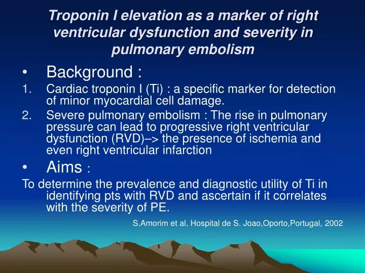
Troponin I elevation as a marker of right ventricular dysfunction and severity in pulmonary embolism. Background : Cardiac troponin I (Ti) : a specific marker for detection of minor myocardial cell damage.

E N D
Troponin I elevation as a marker of right ventricular dysfunction and severity in pulmonary embolism • Background : • Cardiac troponin I (Ti) : a specific marker for detection of minor myocardial cell damage. • Severe pulmonary embolism : The rise in pulmonary pressure can lead to progressive right ventricular dysfunction (RVD)–> the presence of ischemia and even right ventricular infarction • Aims : To determine the prevalence and diagnostic utility of Ti in identifying pts with RVD and ascertain if it correlates with the severity of PE. S.Amorim et al, Hospital de S. Joao,Oporto,Portugal, 2002
Methods • 77 pts with PE • Classified the PE in severity levels (by ESC) 1-massive : shock and /or hypotension 2-submassive : RV hypoplasia found by echo 3-nonmassive : the remaining cases 3. The highest Ti serum value : since the admission until 24 hours and a normal value of <0.10 ng/ml
Results • 60 pts with Ti measurements : 42 (elevated Ti values) • Pts with RVD : 26 (81.3%) had increased Ti levels without RVD : only 14 (35%) with elevated Ti level positive Ti test : significantly associated with RVD (P= 0.038) • Positive Ti test : earlier beginning of symptoms (92.5+- 152.79 vs 233.4+-232.47 hours , p=0.02) • Positive Ti test : a higher prevalence of emboli in proximal vessels( pulmonary trunk and right or left pulmonary trunk) (92% vs 52%, p=0.012)
Results-2 • The mean level of Ti : pts with severe PE (1.65+-4.27ng/ml) vs submassive PE(1.06+-0.97ng/ml) vs nonmassive PE(0.53+-0.74ng/ml) (P=0.045) • The level of d-dimers or systolic pressure of pulmonary artery : no gradual relationship between the classes of PE.
Conclusions • Around 55% of pts with PE have elevated Ti. • Ti : significantly associated with RVD • Ti : identification of pts of greater severity and at increased risk of hemodynamic deterioration, which can benefit of more aggressive therapeutical strategies
Determinants of the expansion of different coronary stents in curved stenotic lesions :an in-vitro experimental study Background: • Coronary stent implantation in angulated vessels is a challenging issue • Currently lack of consensus regarding the type and length of stents best suitable for such lesion Aims : To assess comparatively the expansion parameters of several new generation stent types in a curved stenotic phantom. T. Poerner et al, University hospital of Manheim;Technical University,institut fur Biomedizinische Technik, Berline,Germany ,2002
Methods • Identical silicon models of 3.2 mm diameter with a 55% concentric elastic stenosis • A number of 5 stents for each length and type: • AVE (Medtronic ) --- 3.5/12, 3.5/18 mm • Penta (Guidant) --- 3.5/13, 3.5/18 mm • BX-Sonic (Cordis) --- 3.5/13, 3.5/18 mm • FlexMaster (Jomed)--- 3.5/12, 3.5/16 mm
Methods-2 3.The forces exerted during ballon inflation (Finfl) and after ballon deflation (Fstent) : continuously registered at a rate of 20 measurements per second using a high sensitive dynamometer 4. MLD (minimal luminal diameter of stent) RLD (reference luminal diameter of stent) BDmin (minimal luminal diameter of inflated ballon) BDref (reference luminal diameter ofinflated ballon) : were determinated by X –ray imaging with direct magnification
Results • All stents: a good wall apposition without deformation of the phantom curvature • The expansion parameters related to the stent length : no significant differences. • The displacement forces on the vessel : within a low range for all stents (higher for AVE and Penta) • Expansion parameters are summarized in the table below.
Conclusions • In this curved elastic stenotic model • AVE : the lowest recoil • AVE & Penta : reach the largest MLD • FlexMaster : the lowest expansion forces • FlexMaster & BX-Sonic : significant lower MLD values