Download
1 / 59

E N D
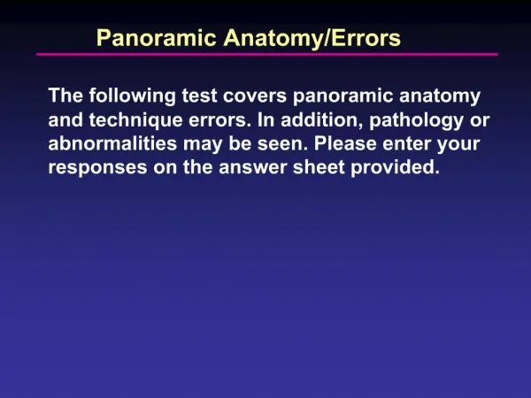
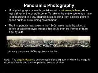
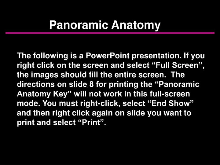
Panoramic Anatomy The following is a PowerPoint presentation. If you right click on the screen and select “Full Screen”, the images should fill the entire screen. The directions on slide 8 for printing the “Panoramic Anatomy Key” will not work in this full-screen mode. You must right-click, select “End Show” and then right click again on slide you want to print and select “Print”.
Types of Panoramic Images Single Real Image Double Real Image Ghost Images
Single Real Image Only one image results from a given anatomical structure. Most images seen on a panoramic film are of this type.
Double Real Image Two images of a single object which is located in the midline. Structures that produce these double real images include the hard & soft palate, hyoid bone and cervical spine.
Ghost Image Usually caused by external objects such as earrings but may be produced by dense anatomical structures such as the mandible
11 16 12 2 5 15 13 17 10 6 7 9 8 14 19 1 24 23 21 18 3 4 32 25 20 26 22 27 31 29 33 37 39 35 30 28 38 36 34
47 40 41 46 42 44 45 43
Panoramic Anatomy Key • 1. maxillary sinus • 2. pterygomaxillary fissure • 3. pterygoid plates • 4. hamulus • 5. zygomatic arch • 6. articular eminence • 7. zygomaticotemporal suture • 8. zygomatic process • 9. external auditory meatus • mastoid process • middle cranial fossa • lateral border of the orbit • infraorbital ridge • infraorbital foramen • infraorbital canal • nasal fossa • nasal septum • anterior nasal spine • inferior concha • incisive foramen • hard palate • maxillary tuberosity • condyle • coronoid process 25. sigmoid notch 26. medial sigmoid depression 27. styloid process 28. cervical vertebrae 29. external oblique ridge 30. mandibular canal 31. mandibular foramen 32. lingula 33. mental foramen 34. submandibular gland fossa 35. internal oblique ridge 36. mental fossa 37. mental ridges 38. genial tubercles 39. hyoid bone 40. tongue 41. soft palate 42. uvula 43. posterior pharyngeal wall 44. ear lobe 45. glossopharyngeal air space 46. nasopharyngeal air space 47. palatoglossal air space You may print this page by right-clicking and selecting “Print”
9 12 7 19 5 17 13 25 14 6 22 18 39 28 33 9 19 7 12 17 13 14 5 6 25 22 18 39 28 33
11 2 15 24 26 32 8 23 16 1 31 3 20 4 34 44 30 38 2 11 15 24 32 26 8 23 16 1 31 3 20 44 34 30 38
46 21 41 42 47 40 45 43 46 21 41 42 47 40 45 43
11 7 1 46 41 47 43 36 45 38
16 17 23 2 8 6 21 18 19 39 Red arrows point to ghost image of hard palate
11 9 3 20
17 2 44 20 28 43
2 atlas 31 transverse foramen
15 46 47 19 6 27 34
17 1 8 15 32 N
40 27 E LN 36
2 8 40 18 45 ? ? Identifies calcification, possibly in carotid or in lymph node
8 7 46 47 33 epiglottis
11 21 3 29 32 34 Red arrow identifies static electricity, caused by removing the film too quickly from the cassette or from the box of film (creates friction, which results in a static discharge).
16 10 9 20 3 42 27 30 1 44 ghost of mandible 36
24 14 27 47 nose 39 Lead apron located too high on back of patient’s neck.
12 air cell 9 23 7 26 Air cell in zygomatic arch.
24 7 26 22 27 30 38
5 10 6 47 45 ghost of mandible
15 23 7 9 21 39 30 Note the relatively inferior location of the mandibular canal (30), providing plenty of room for the implant.
24 26 31 1 29 Pattern on right side of film (patient’s left) caused by excessive oil on patient’s hair.
7 28 28 red arrow identifies fracture
27 44 34 Green arrow identifies “pseudo-fracture” caused by palatoglossal air space. Red arrows point to odontogenic keratocyst.
27 28 28 Hearing aid (red arrow) with ghost (green arrow).
Ghost images of mandibles (dotted line outlines ghost of left ramus-angle over right side of mandible)
Slide # 1 C E D G F B A Answers on next slide
Slide # 1 C E D G F B A A = cervical vertebra; B = external oblique ridge; C = zygomatic process; D = floor of maxillary sinus; E = zygomaticotemporal suture; F = lingula; G = mandibular foramen
Slide # 2 B K D J E I A H F C G Answers on next slide
Slide # 2 B K D J E I A H F C G A = ear; B = external auditory meatus; C = submandibular fossa; D = nasal septum; E = hard palate; F = mental foramen; G = hyoid bone; H = mandibular canal; I = pterygoid plates; J = articular eminence; K = pterygomaxillary fissure
Slide # 3 C B D A E Answers on next slide
Slide # 3 C B D A E A = palatoglossal air space; B = middle cranial fossa; C = lateral border of orbit; D = condyle; E = mental fossa
Slide # 4 I D E H C B A G F J K L Answers on next slide
Slide # 4 I D E H C B A G F J K L A = cervical vertebra; B = zygomaticotemporal suture; C = zygomatic process; D = nasal septum; E = inferior concha; F = soft tissue of nose; G = hard palate; H = posterior border of maxillary sinus; I = external auditory meatus; J = posterior pharyngeal wall; K = mental foramen; L = mental fossa
Slide # 5 E F G C D J H B I A Answers on next slide
Slide # 5 E F G C D J H B I A A = glossopharyngeal air space; B = styloid process; C = nasopharyngeal air space; D = pterygoid plates; E = condyle; F = infraorbital canal; G = infraorbital foramen; H = soft palate; I = mandibular canal; J = lingula
Slide # 6 E C D E B F G A Answers on next slide
Slide # 6 E C D E B F G A A = mental foramen; B = incisive foramen; C = soft tissue of nose; D = anterior nasal spine; E = pterygoid plates; F = ear; G = hyoid bone The radiolucency (red arrows) seen in the ramus and third molar area on the patient’s right side is an ameloblastoma. (Differential includes dentigerous cyst, radicular cyst, OKC).
Slide # 7 A B C D Answers on next slide