Download
1 / 27
280 likes | 667 Vues
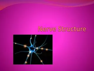
Nerve Tissue. Al- Maarefa College. Objective. Understand the microscopic difference between excitable and non-excitable cells present in the nervous system. Nerve Tissue. Cells have very high ability to Respond to stimuli Transmit impulses. Nerve Tissue. Nerve Tissue.

E N D
Nerve Tissue Al-Maarefa College
Objective • Understand the microscopic difference between excitable and non-excitable cells present in the nervous system.
Nerve Tissue • Cells have very high ability to • Respond to stimuli • Transmit impulses
Nerve Tissue • NEURON is the main nerve cell)
Nerve Tissue • NEURON is the main nerve cell • Cell Body(3) • Dendrites (5) • Axon(1)
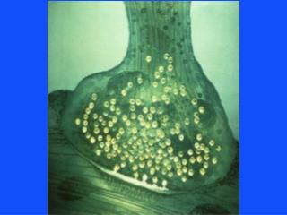
Motor Neurone • Figure 9–2. Motor neuron. The myelin sheath is produced by oligodendrocytes in the central nervous system and by Schwann cells in the peripheral nervous system. The neuronal cell body has an unusually large, euchromatic nucleus with a well-developed nucleolus. The perikaryon contains Nissl bodies, which are also found in large dendrites. An axon from another neuron is shown at upper right. It has 3 end bulbs, one of which forms a synapse with the neuron. Note also the 3 motor end-plates, which transmit the nerve impulse to striated skeletal muscle fibers. Arrows show the direction of the nerve impulse.
Motor Neurone • Cell body • Dendrites • Axon • Covered by Myelin sheath Figure 9–2. Motor neuron. The myelin sheath is produced by oligodendrocytes in the central nervous system and by Schwann cells in the peripheral nervous system. The neuronal cell body has an unusually large, euchromatic nucleus with a well-developed nucleolus. The perikaryon contains Nissl bodies, which are also found in large dendrites. An axon from another neuron is shown at upper right. It has 3 end bulbs, one of which forms a synapse with the neuron. Note also the 3 motor end-plates, which transmit the nerve impulse to striated skeletal muscle fibers. Arrows show the direction of the nerve impulse.
Neurones Figure 9–4. Simplified view of the three main types of neurons, according to their morphological characteristics.
Neuron • Multipolar Kuehnel, Color Atlas of Cytology, Histology, and Microscopic Anatomy
Neuron's – cerebral cortex Kuehnel, Color Atlas of Cytology, Histology, and Microscopic Anatomy
Neuron's – spinal cord Kuehnel, Color Atlas of Cytology, Histology, and Microscopic Anatomy
Nerve and Reflex Arc Figure 9–32. Schematic representation of a nerve and a reflex arc. In this example, the sensory stimulus starts in the skin and passes to the spinal cord via the dorsal root ganglion. The sensory stimulus is transmitted to an interneuron that activates a motor neuron that innervates skeletal muscle. Examples of the operation of this reflex are withdrawal of the finger from a hot surface and the knee-jerk reflex. (Modified, redrawn, and reproduced, with permission, from Ham AW: Histology, 6th ed. Lippincott, 1969.)
Nerve Fiber (Axon) • Nerve fibers are long nerve cell processes (axon cylinder, axon) with a surrounding membrane • Schwann cells (neurolemmocytes, peripheral glial cells) enfold the axon and form an insulating cover known as Schwann’s sheath (neurolemma). Stain: osmium tetroxide; magnification: × 1000 Kuehnel, Color Atlas of Cytology, Histology, and Microscopic Anatomy
Nerve Fiber (Axon) • Myelinated nerve fiber, i.e., the axon is covered by a myelin sheath, which is rich in lipids. • Every 0.8 to 1.0 mm, a node of Ranvier subdivides the myelin sheath into segments or internodes. Stain: osmium tetroxide; magnification: × 1000 Kuehnel, Color Atlas of Cytology, Histology, and Microscopic Anatomy
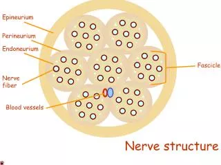
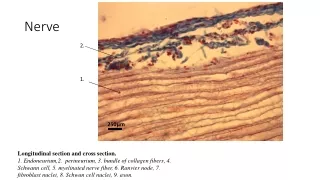
Sciatic nerve – cross section • 1 Fascicle (nerve fiber bundle) • 2 Perineurium • 3 Epineurium • 4 Artery • 5 Vein • 6 Adipose tissue Stain: alum hematoxylin; magnification: × 10 Kuehnel, Color Atlas of Cytology, Histology, and Microscopic Anatomy
Glial Cells • Glial cells (Neuroglia) or (Glia) • (Greek "glue” • Non-neuronal cells that maintain homeostasis, form myelin, and provide support and protection for the brain's neurons • They occupy the entire space between neurons and separate nerve cells from blood vessels
Glial Cells - Functions • Surround neurons and hold them in place • Supply nutrients and oxygen to neurons • Insulate one neuron from another • Destroy pathogens and remove dead neurons
Glial Cells - Types • Astrocytes (macroglia) • Oligodendrocytes • Microgliocytes
Astrocytes • Most abundant • Deal wit homeostasis – relate to vessels Kuehnel, Color Atlas of Cytology, Histology, and Microscopic Anatomy Figure 9–13. Drawings of neuroglial cells as seen in slides stained by metallic impregnation. Note that only astrocytes exhibit vascular end-feet, which cover the walls of blood capillaries.
Astrocytes and nerve cells Kuehnel, Color Atlas of Cytology, Histology, and Microscopic Anatomy
Microglia • Figure 9–13. Drawings of neuroglial cells as seen in slides stained by metallic impregnation
Microglia • Oligodendrocytes: • Closely related to neurons • Provide myelin protection for CNS neurons Figure 9–13. Drawings of neuroglial cells as seen in slides stained by metallic impregnation
Neurons and glial cells Kuehnel, Color Atlas of Cytology, Histology, and Microscopic Anatomy
Neuron and glial cells Kuehnel, Color Atlas of Cytology, Histology, and Microscopic Anatomy
Summary • Nerve Tissue Cells: • Neurons: • Myelinated • Non-myelinated
Summary • Nerve Tissue Cells: • Neurons: • Myelinated • Non-myelintaed • Neurolial cells: • Astrocytes • Microglia • Oligodendrocytes