Download
1 / 42
460 likes | 1.11k Vues
Introduction to the Nervous System and Nerve Tissue. Three Basic Functions Sensory Functions: Sensory receptors detect both internal and external stimuli. Functional unit: Sensory or A fferent Neurons

E N D
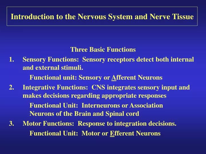
Introduction to the Nervous System and Nerve Tissue Three Basic Functions Sensory Functions: Sensory receptors detect both internal and external stimuli. Functional unit: Sensory or Afferent Neurons Integrative Functions: CNS integrates sensory input and makes decisions regarding appropriate responses Functional Unit: Interneurons or Association Neurons of the Brain and Spinal cord Motor Functions: Response to integration decisions. Functional Unit: Motor or Efferent Neurons
Organization of the Nervous System to supply the three basic functions
Organization of the CNS Gray Matter: Contains neuron cell bodies White Matter: Contains cell extensions organized into tracts W
Introduction to the Nervous System and Nerve Tissue Dendrites: Carry nerve impulses toward cell body. Receive stimuli from synapses or sensory receptors. Cell Body: Contains nucleus and nissl bodies, a form of rough endoplasmic reticulum. Axon: Carry nerve Impulses away from the cell bodies. Axons interact with muscle, glands, or other neurons. Structure of a Neuron
Introduction to the Nervous System and Nerve Tissue Types of Neurons
Introduction to the Nervous System and Nerve Tissue Types of Interneurons
Introduction to the Nervous System and Nerve Tissue 1. Schwann cells that form the myelin sheath Types of Supportive Cells of the PNS
Introduction to the Nervous System and Nerve Tissue 1. Schwann cells that form the myelin sheath Types of Supportive Cells of the PNS
Introduction to the Nervous System and Nerve Tissue 1. Satellite cells associated with sensory neuron cell bodies Types of Supportive Cells of the PNS
Introduction to the Nervous System and Nerve Tissue 1. Satellite cells associated with sensory neuron cell bodies Types of Supportive Cells of the PNS
Introduction to the Nervous System and Nerve Tissue 1. Oligodendrocytes: form the myelin sheath of the CNS Types of Supportive Cells of the CNS (Neuroglia)
Introduction to the Nervous System and Nerve Tissue 2. Astrocytes: Help form the blood-brain barrier, support the appropriate chemical environment for neurons. Types of Supportive Cells of the CNS (Neuroglia)
Introduction to the Nervous System and Nerve Tissue 3. Microglia: Phagocytes in the CNS that engulf microbes and cellular debris. Types of Supportive Cells of the CNS (Neuroglia)
Introduction to the Nervous System and Nerve Tissue 4. Ependymal Cells: Form blood-brain barrier in the brain ventricles and central canal of spinal cord. Produce cerebrospinal fluid and assist in its circulation. Types of Supportive Cells of the CNS (Neuroglia)
Nervous System Physiology: Distribution of Ions between ECF and ICF
Nervous System Physiology:Nerve Conduction Occurs because of Changes in Membrane Potential
Nervous System Physiology:Mechanism that creates an Action Potential
Nervous System Physiology:Two Mechanisms of Action Potential Conduction along a neuron
Types of Nerve Fibers • “A” fibers: Largest diameter myelinated fibers with the fastest saltatory conduction (12-130 m/sec) and a brief absolute refractory period. Axons of motor neurons and axons of sensory neurons that conduct touch, pressure, and thermal sensations. (GSSN) • “B” fibers: intermediate diameter myelinated fibers With slower saltatory conduction then “A” fibers and longer absolute refractory periods. Dendrites of visceral sensory neurons and axons of presynaptic neurons of the ANS.
Types of Nerve Fibers • “C” fibers: Smallest diameter unmyelinated fibers with slow continuous conduction (.5 – 2 m/sec.) and the longest absolute refractory periods. Axons of some somatic sensory neuron that carry pain, touch, pressure and thermal sensation, neuron that carry visceral pain sensations, and postsynaptic neurons of the ANS
Nervous System Physiology: Communication between neurons at a synaptic junction 1. Electrical Synapses: Communication via gap junctions between smooth muscle, cardiac muscle, and some neurons of the CNS. Provide fast, synchronized, and two-way transmission of information. 2. Chemical Synapses: Communication via chemical neurotransmitters that diffuse across a synaptic cleft. Provides slow one-way information flow
Nervous System Physiology: Communication between neurons at a synaptic junction • Action potential arrives at • a synaptic end bulb. • Depolarization of membrane • causes the opening of Ca2+ • channels. • Increase in (Ca2+) inside of • presynaptic neuron triggers • exocytosis of neurotransmitter • Neurotransmitter diffuses across • synaptic cleft and binds to • receptor (ligand-gated channel) • on postsynaptic neuron
Nervous System Physiology: Communication between neurons at a synaptic junction • Na+ channels open causing a depolarization (Na+ channels) • EPSP (excitatory postsynaptic potential) or a hyperpolarization (Cl- • channels) IPSP (inhibitory post- • synaptic potential) of the postsynaptic neuron. • If depolarization reaches a threshold, an action potential is generated on the postsynaptic • neuron.
Nervous System Physiology: Communication between neurons at a synaptic junction
Nervous System Physiology: Communication between neurons at a synaptic junction • Neurotransmitters • Acetylcholine: Found in the PNS and CNS. EPSP and in parasympathetic neurons IPSP. • Amino Acids: Glutamate and Aspartate produce EPSP’s in the CNS. Gamma Aminobutyric Acid (GABA) produces IPSP’s in the CNS. Valium enhances the action of GABA.
Nervous System Physiology: Communication between neurons at a synaptic junction • Neurotransmitters • Biogenic Amines: Norepinephrine and epinephrine produce EPSP’s in the sympathetic system. Serotonin controls mood and induction of sleep. • Gases: Nitric Oxide produce by the enzyme nitric oxide synthase. Causes vasodilation and erection.
Nervous System Physiology: Communication between neurons at a synaptic junction • Neurotransmitters • Neuropeptides: • Substance P: Enhances perception of pain. • Endorphins: inhibit pain by blocking release of Substance P • ATP-Adenosine 5’-triphosphate • Between taste buds and nerves that carry taste sensations –Finer et. al. Science vol 310, 2005
Nervous System Physiology: Communication between neurons at a synaptic junction
Brain Waves • Alpha waves: (8 – 13 Hz) Occur when a person is awake, resting, mind wandering and eyes closed. Recorded in the parieto-occipital area. • Beta waves: (14 -30 Hz) Become accentuated during mental activity and sensory stimulation. Recorded in the frontal to parietal regions.
Brain Waves • Theta waves: (4 -7 Hz) Normal in children and drowsy or sleeping adults. Predominant waves in awake adults suggest emotional stress or brain disorders. • Delta waves: (< 3.5 Hz) High-amplitude wave. Infants exhibit these waves when awake and adults exhibit them in deep sleep. Increased delta waves in awake adults indicate serious brain damage.