Download
1 / 30
310 likes | 510 Vues
11. P A R T A. Fundamentals of the Nervous System and Nervous Tissue. Nervous System. The master controlling and communicating system of the body Functions Sensory input – monitoring stimuli Integration – interpretation of sensory input Motor output – response to stimuli. Nervous System.

E N D
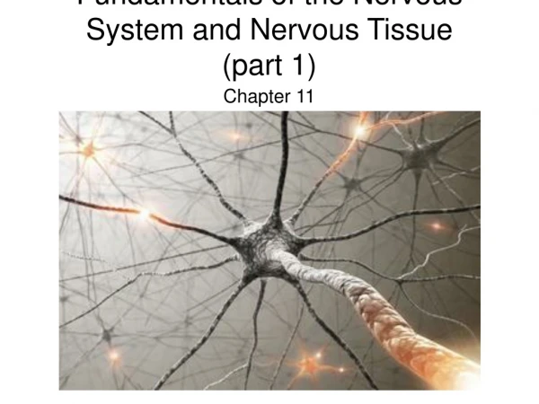
11 P A R T A Fundamentals of the Nervous System and Nervous Tissue
Nervous System • The master controlling and communicating system of the body • Functions • Sensory input – monitoring stimuli • Integration – interpretation of sensory input • Motor output – response to stimuli
Nervous System Figure 11.1
Organization of the Nervous System • Central nervous system (CNS) • Brain and spinal cord • Integration and command center • Peripheral nervous system (PNS) • Paired spinal and cranial nerves • Carries messages to and from the spinal cord and brain
Peripheral Nervous System (PNS): Two Functional Divisions • Sensory (afferent) division • Sensory afferent fibers – carry impulses from skin, skeletal muscles, and joints to the brain • Visceral afferent fibers – transmit impulses from visceral organs to the brain • Motor (efferent) division • Transmits impulses from the CNS to effector organs
Motor Division: Two Main Parts • Somatic nervous system • Conscious control of skeletal muscles • Autonomic nervous system (ANS) • Regulates smooth muscle, cardiac muscle, and glands • Divisions – sympathetic and parasympathetic
Histology of Nerve Tissue • The two principal cell types of the nervous system are: • Neurons – excitable cells that transmit electrical signals • Supporting cells – cells that surround and wrap neurons
Supporting Cells: Neuroglia • The supporting cells (neuroglia or glial cells): • Provide a supportive scaffolding for neurons • Segregate and insulate neurons • Guide young neurons to the proper connections • Promote health and growth
Astrocytes • Most abundant, versatile, and highly branched glial cells • They cling to neurons and their synaptic endings, and cover capillaries
Astrocytes • Functionally, they: • Support and brace neurons • Anchor neurons to their nutrient supplies • Guide migration of young neurons • Control the chemical environment
Astrocytes Figure 11.3a
Oligodendrocytes, Schwann Cells, and Satellite Cells • Oligodendrocytes – branched cells that wrap CNS nerve fibers • Schwann cells (neurolemmocytes) – surround fibers of the PNS • Satellite cells surround neuron cell bodies with ganglia
Oligodendrocytes, Schwann Cells, and Satellite Cells Figure 11.3d, e
Neurons (Nerve Cells) • Structural units of the nervous system • Composed of a body, axon, and dendrites • Long-lived, amitotic, and have a high metabolic rate • Their plasma membrane function in: • Electrical signaling • Cell-to-cell signaling during development
Neurons (Nerve Cells) Figure 11.4b
Nerve Cell Body (or Soma) • Contains the nucleus and a nucleolus • Is the major biosynthetic center • Is the focal point for the outgrowth of neuronal processes • Has no centrioles (hence its amitotic nature) • Contains an axon hillock – cone-shaped area from which axons arise
Processes • Armlike extensions from the soma • Called tracts in the CNS and nerves in the PNS • There are two types: axons and dendrites
Dendrites of Motor Neurons: Structure and Function • Short, tapering, and diffusely branched processes • They are the receptive, or input, regions of the neuron
Axons: Structure and Function • Slender processes of uniform diameter arising from the hillock • Long axons are called nerve fibers • Usually there is only one unbranched axon per neuron • Rare branches, if present, are called axon collaterals • Axonal terminal – branched terminus of an axon • Generate and transmit action potentials • Secrete neurotransmitters from the axonal terminals
Myelin Sheath • Whitish, fatty (protein-lipoid), segmented sheath around most long axons • Formed by Schwann cells in the PNS • Formed by oligodendrocytes in CNS • It functions to: • Protect the axon • Electrically insulate fibers from one another • Increase the speed of nerve impulse transmission
Myelin Sheath: Formation Figure 11.5a–c
Nodes of Ranvier (Neurofibral Nodes) • Gaps in the myelin sheath between adjacent Schwann cells
Axons of the CNS • Both myelinated and unmyelinated fibers are present • Myelin sheaths are formed by oligodendrocytes
Regions of the Brain and Spinal Cord • White matter – dense collections of myelinated fibers • Gray matter – mostly soma and unmyelinated fibers
Neuron Classification • Functional: • Sensory (afferent) — transmit impulses toward the CNS • Motor (efferent) — carry impulses away from the CNS • Interneurons (association neurons) — shuttle signals through CNS pathways
Conduction Velocities of Axons • Conduction velocities vary widely among neurons • Rate of impulse propagation is determined by: • Axon diameter – the larger the diameter, the faster the impulse • Presence of a myelin sheath – myelination dramatically increases impulse speed
Synapses • A junction that mediates information transfer from one neuron: • To another neuron • To an effector cell • Presynaptic neuron – conducts impulses toward the synapse • Postsynaptic neuron – transmits impulses away from the synapse • Are electrical and chemical synapses (the later uses neurotransmitters)
Synapses Figure 11.17
Synaptic Cleft • Fluid-filled space separating the presynaptic and postsynaptic neurons • Prevents nerve impulses from directly passing from one neuron to the next • Transmission across the synaptic cleft: • Is a chemical event (as opposed to an electrical one) • Ensures unidirectional communication between neurons
Neurotransmitters (write in notes) • Chemicals used for neuronal communication with the body and the brain • 50 different neurotransmitters have been identified • Classified chemically and functionally