Download
1 / 33
330 likes | 581 Vues
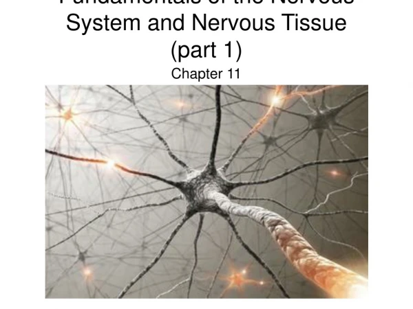
Fundamentals of the Nervous System and Nervous Tissue. Classification. Functions of the Nervous System Organization of the CNS and PNS. The Nervous System. The Nervous System is the rapid control system of the body There are two anatomical divisions to the Nervous System:

E N D
Classification Functions of the Nervous System Organization of the CNS and PNS
The Nervous System • The Nervous System is the rapid control system of the body • There are two anatomical divisions to the Nervous System: • The Central Nervous System (CNS) • The Peripheral Nervous System (PNS) • They work together as a single coordinated whole
The Functions of the Nervous System • There are three interconnected functions: • Sensory input • from millions of specialized receptors receive stimuli • Integration • process stimuli and interpret stimuli • Motor output • cause response • at many effector organs
Organization of the CNS • The brain and spinal cord • Process & integrate information, store information, determine emotions • Initiate commands for muscle contraction, glandular secretion and hormone release (regulate and maintain homeostasis) • Connected to all other parts of the body by the Peripheral Nervous System (PNS)
Organization of the PNS • Anatomical connections • spinal nerves are connected to the spinal cord • cranial nerves are connected to the brain • Two functional subdivisions • Sensory (afferent) division • somatic afferents - skin, skeletal muscle, tendons, joints • special sensory afferents • visceral afferents - visceral organs • Motor (efferent) division • motor (efferent) neurons • muscles/glands
Organization of the PNS • Motor (efferent) division has two parts: • Somatic Nervous System (SNS) • voluntary motor neurons • output to skeletal muscles • Autonomic Nervous System (ANS) • involuntary visceral motor neurons • output to smooth muscle, cardiac muscles and to glands • two cooperative components: • sympathetic division • parasympathetic division
Autonomic Nervous System • Sympathetic Division – for muscular exertion and for “fight or flight” emergencies • Parasympathetic Division – for metabolic/ physiologic activities; “resting and digesting”
Histology Neuroglia Structure of the Neuron Neuron Classification Nerves
Histology of Nervous Tissue • Despite the complexity of organization, there are only two functional cell types • neuroglia (glial) cells - support cells • neurons (nerve cells)- excitable nerve cells that transmit electrical signals (messages) from one part of the body to another
Histology of CNS Tissue - Neuroglia • Do not conduct nerve impulses • Divide throughout life. Good: repair themselves Bad: source of most brain tumors (gliomas). • 4 types of glial cells in CNS and 2 types in PNS
Histology of CNS Tissue – Neuroglia • Astrocytes • star shaped with many processes • connect to neurons; help anchor them to nearby blood capillaries (part of blood-brain barrier) • control the chemical environment of the neurons; recapture neurotransmitters • Microglia • oval with thorny projections • monitor the health of neurons; if infection occurs, they change into macrophages (eating viruses, bacteria and damaged cells)
Histology of CNS Tissue - Neuroglia • Ependymal cells • range in shape from squamous to columnar; many are ciliated; help circulate cerebrospinal fluid • line the central cavities of the brain and spinal cord • form a protective cushion around the CNS (brain and spinal cord) • Oligodendrocytes • line up along neurons and wrap themselves around axons • form the myelin sheath – a fatty, insulating membrane
Histology of PNS Tissue - Neuroglia • Satellite cells • surround neuron cell bodies in the PNS • Protective, cushioning cells • Schwann cells (neurolemmocytes) • surround axons/dendrites and form the myelin sheath around larger nerve fibers in the PNS • similar to oligodendrocytes in function - insulators
Histology of CNS Tissue - Neurons • Neurons - highly specialized cells which conduct electrochemical signals (nerve impulses) • Extreme longevity – neurons live and function normally for a lifetime • Amitotic • once mature, neurons lose the ability to divide • damaged nervous tissue cannot regenerate • High metabolic rate • need a large, constant supply of oxygen and glucose • can survive only a few minutes without oxygen
Neuron Structure – Cell Body • Contains the usual cellular organelles • Synthesis & metabolism occurs primarily in the cell body • The outer cell membrane contains the receptors for incoming information (stimuli) • Most cell bodies are located within the CNS • clusters of neuron cell bodies in the CNS are usually called nuclei • clusters of neuron cell bodies in the PNS are usually called ganglia
Neuron Structure – Processes (fibers) • Dendrites • short, tapering, highly branched processes or extensions from the cell body • transfer information to the cell body • Neurons may have hundreds of branching dendrites depending on the neuron type
Neuron Structure – Processes (fibers) • Axons • a long thin cylindrical cytoplasmic projection • starts at a cone-shaped region – the axon hillock • transmit information away from the cell body • ends in many branches • known as axonal terminals • may be 10,000 on one nerve • form synapses (junctions) with neighboring neurons or with effector cells (muscles or glands) *Each neuron only has one axon
Neuron Structure – Processes (fibers) Axons - continued • Nerve impulses start at the axon hillock; travel along the axon to the axon terminal • axon terminal has the secretory component • Action Potential (nerve impulse) arrival causes the release of stored neurotransmitters from their vesicles • Neurotransmitters transfer the message across a synapse to the next neuron or to an effector cell • Neurotransmitters can excite or inhibit the action of the next cell in the pathway
Each axon terminal is separated from the next neuron by a tiny gap called the synaptic cleft • Although they are close, neurons never actually touch other neurons
Neuron Structure – Myelin Sheath • Myelin sheath • White, lipid-rich, segmented covering on an axon • primarily along the larger, longer axons • dendrites are never myelinated • myelin protects & electrically insulates the axon • increases the speed of nerve impulses • myelinated fibers conduct impulses 10-150x faster than unmyelinated fibers • 150 m/sec vs. 1 m/sec
Myelinating Cells • Schwann cells (neurolemmocytes) in the Peripheral NS • Oligodendrocytes in the Central NS
Myelination • Occurs during fetal development and the first year of life • Each myelinating cell wraps around an axon up to 100 times • myelin sheath: multiple layers of the cell membrane • neurilemma (sheath of Schwann): outer layer containing the bulk of the cytoplasm and cell organelles; can stay intact in PNS damage – aids in regeneration after an injury
Myelinated and Unmyelinated Axons • Myelinated Fibers • Has Myelin sheaths and neurofibril nodes (nodes of Ranvier) - periodic gaps every mm in the myelin sheath between the neurolemmocytes • Unmyelinated Fibers • the axon rest in the indentations of the oligodendrocytes or the Schwann cells; no myelin sheath present
Neuron Structure – Processes (fibers) • Fibers can be sensory or motor fibers • Groups of fibers in the CNS are called tracts; groups of fibers in the PNS are called nerves
Gray Matter vs. White Matter (CNS) • Gray matter - unmyelinated cell bodies & processes (tracts), ex. Cerebral cortex and basal nuclei in brain and core of spinal cord • White matter – myelinated processes in various fiber tracts, ex. Cerebral white matter of brain and outer part of spinal cord
Neuron Classification • Functional: based on the direction (location) of nerve impulses; we emphasize the functional classification > Afferent (sensory) neurons > Efferent (motor) neurons > Interneurons Ratios: 1 afferent : 10 efferent : 200,000 interneurons
Afferent (Sensory) Neurons • Nerve impulses from specific sensory receptors (touch, sight, etc.) are transmitted to the spinal cord or brain (CNS) • Keeps us informed about what is happening inside and outside the body • Afferent neuron cell bodies are located outside the CNS in the ganglia of the PNS
Efferent (Motor) Neurons • Nerve impulses from CNS (brain and spinal cord) are transmitted to effectors (muscles, endocrine and exocrine glands) • Efferent neuron cell bodies are located inside the CNS
Association Neurons (Interneurons) • Carry nerve impulses from one neuron to another • They connect the motor and sensory neurosn in neural pathways • 90% of the neurons in the body are interneurons • Most interneurons are located in the CNS
Neuron Classification • Structural: based on the number of processes extending from the cell body > Multipolar – several dendrites and one axon (ex. Motor neurons of brain and spinal cord) > Bipolar – 1 axon and 1 dendrite (ex. Receptors of the eye and inner ear) > Unipolar – 1 process extending from the cell body (ex. Always sensory neurons)