Download
1 / 55
580 likes | 857 Vues
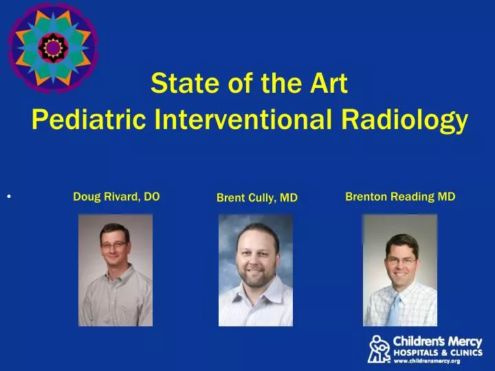
State of the Art Pediatric Interventional Radiology Brent Cully, MD. Doug Rivard , DO Brenton Reading MD. CMH Interventional Radiology. 3 Physicians 2 Nurse Practitioners 3 Technologists 2 IR Rooms 1 CT Fluoro Dedicated Ultrasound. CMH Interventional Radiology.

E N D
State of the ArtPediatric Interventional RadiologyBrent Cully, MD • Doug Rivard, DOBrenton Reading MD
CMH Interventional Radiology • 3 Physicians • 2 Nurse Practitioners • 3 Technologists • 2 IR Rooms • 1 CT Fluoro • Dedicated Ultrasound
CMH Interventional Radiology • Full sedation team under supervision of Dept of Anesthesiology
Sedation • 8 Work Up / Recovery rooms • Most patients get sedation • Must be npo 6 hours • 2 hrs clears
CMH Interventional Radiology • Services Provided • Vascular Access • Angiography / Angioplasty • GI Access • Lumbar Puncture • Image-Guided Biopsy / Drainage • Ablation / Embolization • Catheter Stripping • Intravascular Foreign Body Retrieval
CMH Interventional Radiology • Services Provided • Vascular Anomalies Clinic • In Conjunction with Dermatology and Plastic Surgery • Contact Dermatology Clinic • Direct Interventional Consults • Imaging Guided Biopsies, Drainages, Vascular Access • Contact CMH Radiology
Vascular Access • Placed 368 PICCs in 2010 • In addition to dedicated Vascular Access Team of 8 nurses • As small as 2.6 Fr DL 109 Port-A-Caths in 2010
Angiography / Angioplasty • 15 year old with recurrent dialysis graft issues
4 yo playing with Mom’s BP cuff • 190 /110 mmHg
6 year old girl s/p liver transplant, now with elevated LFTs and splenomegaly
CT angio shows stenosis at the portal vein anastomosis • Post-stenotic dilation of the intrahepatic portal vein • Dilated intrahepatic bile ducts
Ultrasound-Guided PercutaneousTranshepaticCholangiogram • Internal / External Biliary Drain
GI Access - Cecostomy • Provide easy colon access for patients needing daily enema therapy Constipation
GI Access – Perc GT and GJ • Initial placement of percutaneous GT tubes • Ultrasound liver margin, contrast enema to outline colon, inflate stomach thru NG
Gastric Port Jejunal Port
Percutaneous Drainage • Percutaneously drained approx 50 periappendiceal abscesses last year • Currently in study of tPA infusion into abscesses to ? decrease hospital stay
Abscess Drainage • 17 yo female treated with 1 month of steroid therapy for inflammatory bowel disease • Developed chest pain and right shoulder pain, fever Pneumonia
CT chest shows a large liver abscess with diaphragm perforation
Placed percutaneous drain with US guidance • Cultures grew Streptococcus anginosus
Percutaneous Drainage • 15 year old who developed fever and cough after visiting her father in Michigan • Positive Histoplasma titers
Sclerotherapy • Imaging – guided injection of lymphatic and venolymphatic malformations for nonsurgical treatment, or size reduction prior to surgery • Irritation of internal lining of the fluid cavity • Resultant scarring, limited re-expansion • Doxycycline, Sotradecol (detergent)
2 year old girl who developed left neck and axilla swelling following URI • Findings consistent with infected or reactive lymphatic malformation
Access obtained with Ultrasound • Contrast injected to assess communication between cavities and ensure no systemic venous runoff • Sclerosant injected, +/- small drain for next few days
Sclerotherapy • Does not completely resolve lesion • Goal is cosmetic improvement, functionality • Will require multiple treatments
Laser Ablation • 2 year old girl with large venous malformation of right leg
Laser catheter introduced into vein lumen • Saline injected around vein to act as heat sink • Laser “fired” and slowly withdrawn
Pre Operative Embolization • 18 year old male with lifelong flank mass, biopsy proven AVM • Requesting excision, surgeon concerned about bleeding