Download
1 / 40
410 likes | 514 Vues
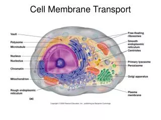
CH. 5 CELL MEMBRANE AND TRANSPORT. A Closer Look at Cell Membranes. BOZEMAN VIDEO—CELL MEMBRANES. http://www.youtube.com/watch?v=y31DlJ6uGgE http://www.youtube.com/watch?v=S7CJ7xZOjm0. http://www.youtube.com/watch?v=y31DlJ6uGgE http://www.youtube.com/watch?v=S7CJ7xZOjm0. Lipid Bilayer.

E N D
CH. 5 CELL MEMBRANE AND TRANSPORT A Closer Look at Cell Membranes
BOZEMAN VIDEO—CELL MEMBRANES http://www.youtube.com/watch?v=y31DlJ6uGgE http://www.youtube.com/watch?v=S7CJ7xZOjm0
http://www.youtube.com/watch?v=y31DlJ6uGgE http://www.youtube.com/watch?v=S7CJ7xZOjm0
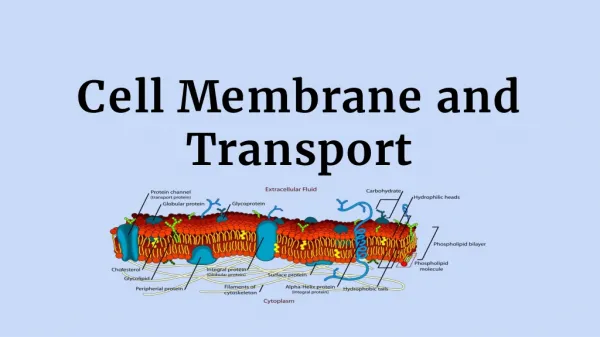
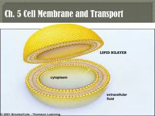
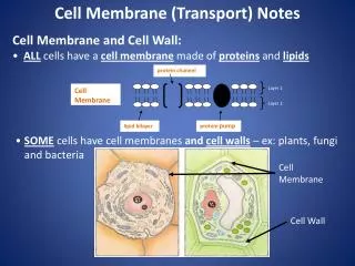
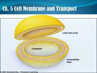
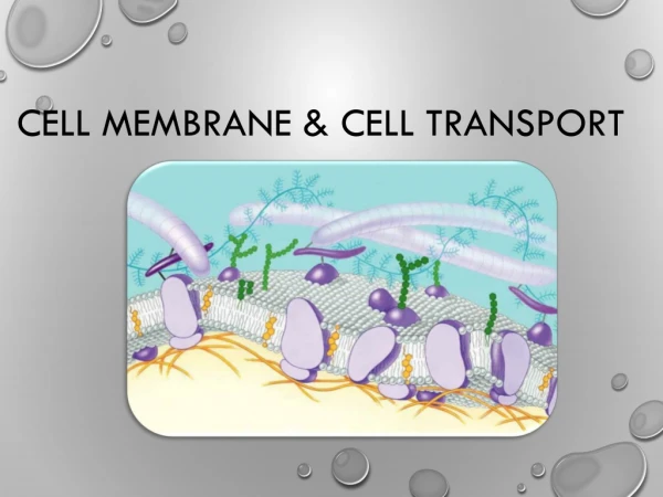
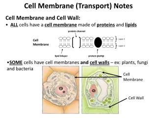
Lipid Bilayer • Cell membranes consist of a lipid bilayer containing different proteins • Membrane is a continuous boundary layer that selectively controls the flow of substances across it
hydrophilic parts hydrophobic parts fluid fluid one layer of lipids one layer of lipids b cross-section through lipid bilayer a Fig. 5.3, pg. 76
Fluid Mosaic Model • Every cell membrane has a mixed composition of phospholipids, glycolipids, sterols, and proteins • KNOW THE CAMPBELL DIAGRAM “CELL MEMBRANE MOSAIC”
Fluid Mosaic Model passive transporter recognition protein phospholipid adhesion protein cholesterol receptor Lipid bilayer active transporter (ATPase pump) active transporter (calcium pump) Cytoplasm Plasma Membrane cytoskeletal proteins just beneath the plasma membrane Fig. 5.4, pg. 77
Studying Membranes Stepped Art Fig. 5.5a, pg. 77
Overview of Membrane Proteins Adhesion Proteins Communication Proteins Fig. 5.6, p.78
Overview of Membrane Proteins Receptor Proteins Recognition Proteins Passive Transporters Active Transporters Fig. 5.6, p.79
Transport Proteins • Span the lipid bilayer • Interior is able to open to both sides • Change shape when they interact with solute • Play roles in active and passive transport
Concentration Gradient • Means the number of molecules or ions in one region is different than the number in another region • In the absence of other forces, a substance moves from a region where it is more concentrated to one one where it’s less concentrated - “down” gradient
Diffusion • The net movement of like molecules or ions down a concentration gradient • Although molecules collide randomly, the net movement is away from the place with the most collisions (down gradient)
Diffusion Stepped Art Fig. 5.7a, p.80
Factors Affecting Diffusion Rate • Steepness of concentration gradient • Steeper gradient, faster diffusion • Molecular size • Smaller molecules, faster diffusion • Temperature • Higher temperature, faster diffusion • Electrical or pressure gradients
Membrane Crossing Mechanisms Diffusion across lipid bilayer Passive transport Active transport Endocytosis Exocytosis
Cell Membranes Show Selective Permeability oxygen, carbon dioxide, and other small, nonpolar molecules; some water molecules glucose and other large, polar, water-soluble molecules; ions (e.g., H+, Na+, K+, Ca++, Cl–); water molecules Fig. 5-8, p.80
Membrane Crossing: Overview I High Concentration gradient across cell membrane ATP Low Diffusion of lipid-soluble Substances across bilayer Passive transport of water- soluble substances through channel protein; no energy input needed Active transport through ATPase; requires energy input from ATP Fig. 5-9, p.81
Passive Transport • Flow of solutes through the interior of passive transport proteins down their concentration gradients • Passive transport proteins allow solutes to move both ways • Does not require any energy input
TYPES OF PASSIVE TRANSPORT • DIFFUSION • FACILITATED DIFFUSION—diffusion that is “helped out” by membrane transport proteins (such as helping larger molecules pass)
Passive Transport glucosetransporter solute (glucose) high low Stepped Art Fig. 5.10, p.80
Active Transport • Net diffusion of solute is against concentration gradient • Transport protein must be activated (requires an integral membrane protein) • ATP gives up phosphate to activate protein • Binding of ATP changes protein shape and affinity for solute
TYPES OF ACTIVE TRANSPORT • SODIUM-POTASSIUM PUMP—3 Na move out and allow 2 K to enter; needs net 1 ATP molecule to dothis • PROTON PUMP– pushes H ions out of the membrane allowing them to eventually diffuse back in; generates ATP for the cell; used in cell respiration This uses ATP synthase enzyme • COTRANSPORT– when molecules travel together across the membrane
Types of active transport Continued • Endocytosis—”engulfing “things into cell membrane; difficult for plants to do Ex: white blood cells engulfing bacteria—to fight infections phagocytosis (large food) pinocytosis (small things) Receptor-mediated endocytosis—use of receptor proteins to help pull things into cell Exocytosis—”spitting” things out of cell
Osmosis • Diffusion of water molecules across a selectively permeable membrane • Direction of net flow is determined by water concentration gradient • Side with the most solute molecules has the lowest water concentration
Osmosis p.84
Tonicity Refers to relative solute concentration of two fluids Hypotonic - having fewer solutes Hypertonic - having more solutes Isotonic - having same amount
Tonicity and Osmosis 2% sucrose solution 1 liter of 10% sucrose solution 1 liter of 2% sucrose solution 1 liter of distilled water Hypotonic Conditions Hypertonic Conditions Isotonic Conditions Fig. 5-13, p.85
Pressure and Osmosis • Hydrostatic pressure • Pressure exerted by fluid on the walls that contain it • The greater the solute concentration of the fluid, the greater the hydrostatic pressure • Osmotic pressure • Amount of pressure necessary to prevent further increase of a solution’s volume
Increase in Fluid Volume first compartment second compartment hypotonic solution hypertonic solution membrane permeable to water but not to solutes fluid volume rises in second compartment Fig. 5.14, p.85
ENDOCYTOSIS AND EXOCYTOSIS VIDEO http://www.youtube.com/watch?v=DuDmvlbpjHQ
Endocytosis and Exocytosis • Exocytosis: A cytoplasmic vesicle fuses with the plasma membrane and contents are released outside the cell 3 types: phagocytosis—engulfs large molecules; difficult for plant cells to do (WHY??) pinocytosis—engulfs small molecules receptor-mediated—uses receptor membrane proteins to pull things in • Endocytosis: A small patch of plasma membrane sinks inward and seals back on itself, forming a vesicle inside the cytoplasm – membrane receptors often mediate this process
Macrophage engulfing Leishmaniamexicana parasite macrophage Fig 5.17, p.87
Phagocytosis bacterium phagocytic vesicle Fig. 5-17b, p.87
Contractile Vacuole contractile vacuole filled contractile vacuole emptied Fig. 5.21, pg. 89
Plasmolysis Plasmolysis
adhesion protein passive transporter recognition protein receptor protein lipid bilayer cytoskeletal proteins cytoplasm active transporter (calcium pump) active transporter (ATPase pump) Fig. 5-19, p.88
BOZEMAN VIDEO—CELL TRANSPORT ACROSS MEMBRANES AND ANIMATION http://www.youtube.com/watch?v=RPAZvs4hvGA http://www.youtube.com/watch?v=RPAZvs4hvGA