Download
1 / 31
380 likes | 728 Vues
Fetal Circulation. Mike Haines, MPH, RRT-NPS, AE-C. Heart Chambers and Valves. Heart Innervation. Heart receives visceral motor innervation Sympathetic (speeds up) Parasympathetic (slows down) . Heart and Lung.

E N D
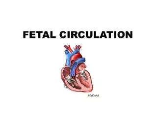
Fetal Circulation Mike Haines, MPH, RRT-NPS, AE-C
Heart Innervation • Heart receives visceral motor innervation • Sympathetic (speeds up) • Parasympathetic (slows down)
Heart and Lung • There are numerous cardiac and pulmonary entities which cause neonatal morbidity and mortality. • Many pulmonary issues (eg. croup, bronchiolitis, pneumonia, etc.) have been discussed previously. • The focus today will be on congenital heart disease.
Heart • Congenital heart disease (CHD) occurs in 1/125 live births. • Neonates may present with a variety of non-specific findings, including: - tachypnea - cyanosis - pallor - lethargy - FTT - sweating with feeds • More specific findings include: - pathological murmurs - hypertension - abnormal pulses - syncope
Neonatal cardiac physiology • The transformation from fetal to neonatal circulation involves two major changes: • A marked increase in systemic resistance. • caused by loss of the low-resistance placenta. 2. A marked decrease in pulmonary resistance. • caused by pulmonary artery dilation with the neonate’s first breaths.
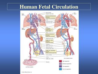
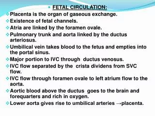
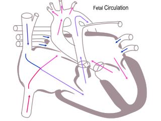
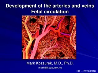
Fetal Circulation • No circulation to lungs • Foramen ovale • Ductus arteriosum • Circulation must go to placenta • Umbilical aa., vv.
Fetal cardiac physiology Fetal circulation: • Blood flows from the placenta IVC RA through the PFO LA LV ascending aorta brain returns via the SVC
Fetal cardiac physiology Fetal circulation: • From the SVC RA RV pulm aa through the PDA descending aorta lower extremities and placenta
Fetal cardiac physiology Fetal circulation: • Only a very small amount of blood is directed through the right and left pulmonary aa’s to the lungs.
Neonatal cardiac physiology Neonate circulation: • The transformation to neonatal circulation occurs with the first few breaths. • The two remaining remnants of the fetal circulation are a patent foramen ovale... and ductus arteriosus.
Congenital Heart Disease • Neonates with CHD often rely on a patent ductus arteriosus and/or foramen ovale to sustain life. • Unfortunately for these neonates, both of these passages begins to close following birth. • The ductus normally closes by 72hrs. • The foramen ovale normally closes by 3 months.
CHD • That being said, in the presence of hypoxia or acidosis (generally present in ductus-dependent lesions), the ductus may remain open for a longer period of time. • As a result, these patients often present to the ED during the first 1-3 weeks of life. • i.e. as the ductus begins to close.
Classifying CHD • There are many different classification systems for CHD. • None are particularly good. • I will be discussing the Pink/Blue/Grey-Baby system: • Pink Baby – Left to right shunt • Blue Baby – Right to left shunt • Grey Baby – LV outflow tract obstruction
Pink Baby (L R shunt) • L R shunts cause CHF and pulmonary hypertension. • This leads to RV enlargement, RV failure, and cor pulmonale. • These babies present with CHF and respiratory distress. • They are not typically cyanotic.
Pink Baby (L R shunt) • These lesions include (among others) ASD’s, VSD’s, and persistently patent ductus arteriosus. VSD ASD
Pink Baby (L R shunt) Persistently patent ductus arteriosus
Pink Baby (L R shunt) • Diagnosing L R shunts depends on: 1. Examination findings: • Non-cyanotic infant in resp distress. • Crackles, widely-fixed second heart sound, elevated JVP, cor pulmonale. 2. CXR: • Increased pulmonary vasculature (suggestive of CHF). • RA and/or RV enlargement.
Pink Baby (L R shunt) • Initial management should be directed at reducing the pulm edema. • Adminster Lasix 1mg/kg IV. • Peds Cardiology/ PICU should be consulted urgently regarding use of: • Morphine • Nitrates • Digoxin • Inotropes
Blue Baby (R L shunt) • R L shunts cause hypoxia and central cyanosis. • Neither hypoxia or cyanosis tend to improve with 100% oxygen. • R L lesions include (among others): • Tetralogy of Fallot (TOF) • Transposition of the Great Arteries (TGA)
Tetralogy of Fallot • Characterized by: • Pulmonary aa OTO • RV hypertrophy • VSD • Over-riding aorta • With severe pulmonary OTO... * * * bloodflow to the lungs may be highly ductus-dependent. *
Tetralogy of Fallot • The classic CXR finding in TOF is the boot-shaped heart. • Pulmonary vasculature is typically decreased.
Transposition of the Great Arteries • TGA is the most common cyanotic lesion presenting in the first week of life. • Anatomically: • RV aorta • LV pulmonary aa • To be compatible with life, mixing of the two circulations must occur via an ASD, VSD, or PDA.
Transposition of the Great Arteries • The CXR findings in TGA are typically less dramatic than in TOF. • Pulmonary vasculature is typically increased.
Blue Baby (R L shunt) • Hypoxia and cyanosis (unresponsive to oxygen) in the neonatal period suggests a ductus-dependent lesion. • Treatment is a prostaglandin-E1 (PGE1) infusion. • Dosing discussed momentarily • This should obviously be accompanied by urgent Peds Cardiology and PICU consultation.
Grey Baby (LVOTO) X • Left-ventricular outflow tract obstructions (LVOTO’s) lead to cyanosis, acidosis, and shock early in the neonatal period. • Complete obstruction is universally fatal unless shunting occurs through an ASD, VSD, or PDA. • Examples of these lesions include: • Severe coarctation of the aorta • Hypoplastic left heart syndrome (HLHS)
Grey Baby (LVOTO) • Treatment: • Any neonate presenting with shock unresponsive to fluids +/- pressors has a LVOTO until proven otherwise. • As with the Blue babies, appropriate management is an urgent PGE1 infusion and emergent consultation.
Prostaglandin-E1 • PGE1 promotes ductus arteriosus patency. • Use an IV infusion at 0.05-0.1 ug/kg/min. • A response should be seen within 15 min. • If ineffective, try doubling the dose. • If effective, try halving the dose. • The lowest possible dose should be used– as adverse-effects of PGE1 can include: - fever - flushing - diarrhea - periodic apnea (be ready to intubate)
Remnants of Fetal Circulation • Ligamentum teres= Round ligament • Remnant of the umbilical vein • Anterior abdominal wall • Ligamentum venosum • Remnant of ductus venosum • On liver’s inferior surface • Medial Umbilical Ligaments • Remnant of umbilical arteries • Anterior abdominal wall below navel • Also gives branch to urinary bladder