Download
1 / 9
90 likes | 197 Vues
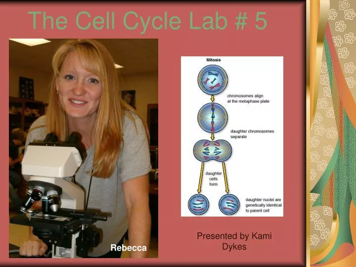
The Cell Cycle Lab # 5. Presented by Kami Dykes. Rebecca. What is each stage of the Cell Cycle?. Interphase. Anaphase. Prophase. Telophase. Metaphase. Cytokinesis. http://www.biology.arizona.edu/cell_bio/tutorials/cell_cycle/cells3.html.

E N D
The Cell Cycle Lab # 5 Presented by Kami Dykes Rebecca
What is each stage of the Cell Cycle? Interphase Anaphase Prophase Telophase Metaphase Cytokinesis http://www.biology.arizona.edu/cell_bio/tutorials/cell_cycle/cells3.html
The ALLIUM (onion root tip) is a region of fast growing tissue where many cells are dividing at any given moment. Furthermore, these plant cells have very nicely defined cell borders (cell walls) and unlike many animal cells are easy to distinguish. http://millette.med.sc.edu/Lab%203%20pages/cell_structure%20II_Lab3.htm
Some of the mitotic figures marked are a. Anaphase b. Prophase c. Interphase a. Anaphase b. Prophase
This is late TELOPHASE at the arrow. Why is this a TELOPHASE figure? In plants, telophase begins the moment you can first detect a cell plate beginning to form, and it does so from the middle or inside out, rather than pinching from the outside inward as in animal cells. This shows a PROPHASE nucleus at (a). What structure do you think is shown at (b)? Can you find another prophase figure? How does it compare with that seen at (a)? Nucleus, the nuclear envelope has dispersed.
This shows an ANAPHASE cell at the arrow. What stage of mitosis is the cell next to it exhibiting? In what stage are most of the cells seen here? Metaphase, prophase
What is each phase of the Blastula (white fish)? http://biog-101-104.bio.cornell.edu/BioG101_104/tutorials/cell_division/wf_review_fs.html PROPHASE is characterized by well-formed chromosomes and a nuclear envelope that is dispersing METAPHASE is characterized by chromosomes lined up in single file in the center of the cell. is characterized by two groups of chromosomes moving towards the opposite poles of the cell. ANAPHASE TELOPHASE is characterized by nuclei that have reached the poles of the cell and the onset of cytokinesis.
Drink with the highest calories per ounce Extreme XXL 45.4 calories/ oz. Jenna “tastes like deodorant”
Can you label the parts? Centriole, Chromatid, Centromere, Spindle fiber, Nuclear envelope