Download
1 / 40
410 likes | 433 Vues
Digestive tract. ---Digestive system: Digestive tract Digestive gland This system is responsible for the mechanical and chemical breakdown of food material, and for absorbing these digestive products into the blood for use as nutrients by the individual cells and tissues of the body.

E N D
---Digestive system: • Digestive tract • Digestive gland This system is responsible for the mechanical and chemical breakdown of food material, and for absorbing these digestive products into the blood for use as nutrients by the individual cells and tissues of the body
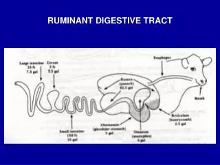
Components of digestive tract ---oral cavity ---pharynx ---esophagus ---stomach ---small intestine ---large intestine
General plan of digestive tract ---Except for oral cavity and pharynx, all other organs share a similar histological plan
General Plan • Mucosa(内膜) • Epithelium • Lamina propria (may contain glands) • Muscularis mucosae (Smooth muscle) • Submucosa(内膜下层) • Loose C.T. may contain glands • Meissner’s autonomic nerve plexus
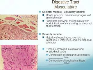
Muscularis externa(肌层) • Inner circular • Myenteric (Auerbach’s) autonomic nerve plexus • Outer longitudinal • Tunica adventitia(外膜) • Fibrosa or serosa (covered by mesothelium)
Esophagus Passage way for food from the pharynx to the stomach mucosa: • epithelium: stratified squamous epithelium • lamina propria: compact CT • muscularis mucosa: longitudinal arranged smooth muscle submucosa: • LCT • esophageal gland: mucous gland
Muscularis externa: • inner circular and outer longitudinal • upper 1/3: skeletal muscle • middle 1/3: mixed of skeletal muscle and smooth muscle • lower 1/3: smooth muscle Tunica adventitia: a fibrous coat of loose connective tissue
Stomach ---dilated part ---store food temporarily ---digest food partially to form a semi-fluid mass, termed chyme ---absorb part of water and ions
Stomach (regions) • Cardia (Cardiac junction) • Surrounds esophageal entrance • Fundic stomach defined histologically includes • Fundus • Body • Pylorus (Pyloric junction) • Pylorus is continuous with the duodenum
Stomach Histology Overview • Mucosa • Epithelium (simple columnar mucus-secreting) • Lamina propria (gastric glands of different types) • Muscularis mucosae (Smooth muscle) • Submucosa • Loose C.T. no glands • Muscularis externa • inner oblique, middle circular, outer longitudinal • Tunica adventitia • Mostly serosa
mucosa • Rugae • Longitudinal folds of mucosa • A mucosal fold contains submucosa • Gastric pits: small depressions, 3-5 gastric gland open into the bottom • Diffuse lymphoid tissue and nodules may be present mucosa
Rugae in the stomach Mucosa Muscularis mucosa Submucosa Muscularis externa Rugae
Cross section of gastric pits Simple columnar epithelium Gastric pit Laminia propria between pits
①epithelium: simple columnar epithelium • surface mucous cell: -tall columnar -ovoid, basally-located nuclei -apical mucin granule -tight junction The mucus is secreted on to the epithelial surface to form a barrier layer which protects it from injury by ingested substance and the stomach’s own secretion of acid and enzymes.
②lamina propria: CT contains fibroblast, LC, plasma cell, mast cell and eosinophil, smooth muscle • gastric gland (fundic gland)-oxyntic gland • cardiac gland: mucous gland • pyloric gland: mucous gland
* Fundic gland -long, branched or unbranched gland
Three part of gland: The neck The body The base Five type cells are found: Chief cells(主细胞) Parietal cells(壁细胞) Mucous neck cells Stem cells Enterendocrine cells neck body
chief cell or zymogenic cell ---structure: LM: • columnar • Round, basally-located Nucleus • cytoplasm: /basal-basophilic /apical-zymogen granules EM: RER, Golgi complex ---function: secret pepsinogen (the precursor of pepsin)
parietal cell or oxyntic cell ---structure: LM: • large, pyramidal or spherical • round centrally-located nucleus • eosinophilic cytoplasm EM: • intracellular secretory canaliculus-invaginations • tubulovesicular system • mitochondria
---function: 1. secret hydrochloric acid (HCl) synthesis processes of HCl: in intracellular secretory canaliculus • H+ K+ -ATP pump: get H+ from cell • Cl- channel: get Cl- from blood • H+ + Cl-→HCl function of HCl: • pepsinogen→pepsin • kill the bacteria
mucous neck cell • less, neck part • pale stain in HE stain • secrete mucus stem cell undifferentiated cell enterendocrine cell • ECL cell: secreting histamine, promote secretion of parietal cell • D cell: secreting somatostatin, inhibit the secretion of parietal cell
Cardiac Junction • Epithelial transition • Stratified Squamous nonkeratinized to simple columnar
Small intestine • Duodenum – first region, only about 25cm long, • Jejunum – second region is roughly 2.5m long • Ileum – last region is roughly 3.5m long • Primary functions • Transport food from stomach to Large intestine • Secretion of digestive enzymes to facilitate digestion of food substances • Absorption of food substances into blood and lymph vessels • Secretion of certain hormones
Small Intestine Overview • Mucosa • Epithelium (simple columnar mucus-secreting) • Lamina propria (intestinal glands) • Muscularis mucosae (Smooth muscle) • Submucosa • loose C.T. (contain duodenal glands in the duodenum) • Muscularis externa • inner circular, outer longitudinal • Tunica adventitia • serosa (except for the duodenum)
Special structure of mucosa • Plicae circulares • Mucosa and submucosa are arranged in permanent, circular mucosal folds • Intestinal villi • Mucosal projections covered by epithelium and containing only lamina propria • Crypt or intestinal glands • Surrounded by lamina propria • Extend to the muscularis mucosae
Plicae circulares Villi
Plicae circulares villi
Epithelium (Simple columnar) • Absorptive cells • Numerous, regular microvilli form striated-border • Well formed junctional complex Plicae circulares, villi and microvilli are serve to increase the surface area of the small intestine by as much as 600-fold surface coat: a layer of glycoprotein filament, protect the underlying cells from mucolytic and proteolytic agent • Goblet cells :secrete mucus to lubricate and protect the epithelium • Enteroendocrine cells: produce hormones
lamina propria: LCT, macrophage, plasma cell and eosinophil and mast cell central lacteal: lymphatic vessel, absorb fat
Crypt or small intestinal gland: the invagination of epithelium into lamina propria • absorptive cell • goblet cell • endocrine cell • stem cell • Peneth cell
Peneth cell: LM: -pyramidal in shape, locate in basal portion of the glands, in groups -apical: acidophilic granules- contain defensin (cryptdin), Lysozyme EM: -protein-secreting cell feature Function: related to immune function, anti-bacterial activity
Large intestine ---Consists of: -cecum -ascending colon -transverse colon -descending colon -sigmoid colon -rectum -anal canal --- function: absorb water and ions
Mucosa • No villi or plicae circulares • Glands are longer than in small intestine • Single columnar epithelium contain numerous goblet cells but absorptive cells are still present • Occasional solitary lymph nodules • At anal junction there is an abrupt transition to stratified squamous non-keratinized epithelium.
Submucosa • Similar to small intestine except nerve plexus are more easily found here • Muscularis externa • Inner circular layer is evident • Outer longitudinal layer forms three bands, the taenia coli • Adventitia • Both serosa and fibrosa are found
Appendix (study by yourself) • Mucosa is like the colon except • Numerous lymph nodules are present in the young • These decrease with age • They break up the muscularis mucosae so that it is difficult to find • The glands are also often not very evident. • Submucosa and muscularis externa are like the rest of the colon