Download
1 / 33
340 likes | 395 Vues
Digestive tract. Dark C. Taste pore. Light C. Taste bud. circumvallated papillae ( HE). filiform and fungiform papillae ( HE). Foliate papillae-taste bud ( HE). Tongue foliate papillae ( HE). Taste pore. ● mucous membrane of tongue

E N D
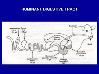
Digestive tract
Dark C Taste pore Light C Taste bud
circumvallated papillae(HE) filiform and fungiform papillae(HE)
Foliate papillae-taste bud (HE) Tongue foliate papillae(HE) Taste pore
● mucous membrane of tongue • Lingual papillae: filiform, fungiform & circumvallated. • Tast bud: dark C, light C & basal C.
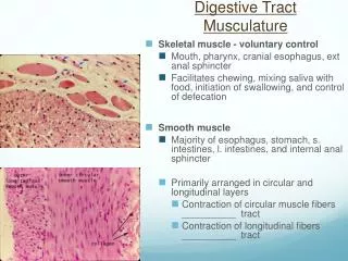
General structure of the wall 1.mucosa: • epithelium: • Lamina propria: • Muscularis mucosa: 2.submucosa: 3.Muscularis externa: 4.adventitia:
4sub mucosa 1Epi. 2lamina propria 4esophageal glands 3 muscula ris mucosa 5inner circular M 6 adven titia 5outer longi tudinal M Esophagus (HE)
epithelium Esophageal gland Esophagus
Esophagus 1.mucosa: ● Epi.: stratified squamous. Non-keratinized ● lamina propria: CT. ● muscularis mucosa: boudles of SMC.longitudinal 2.sbumucosa: esophageal glands, mucous. 3.muscularis: inner circular, outer longitudinal 4.adventitia:fibrosa. • upper1/3:skeletal M. • middle: mixed. • Lower1/3: smooth M.
Stomach • mucosa: • Simple columnar Epi.:surface mucous cell • Lamina propria:gastric pit, gastric glands • Muscularis mucosa: inner circular, outer longitudinal. • submucosa: LCT. • muscularis:inner oblique, middle circular, outer longitudinal. • Adventitia: serosa.
fundic glands location:lamina propria compositions: 1.Chief C 2.Parietal C 3.Neck mucous C 4.Endocrine C 5.Stem C
Secretory granule nucleus rER Chief cells • LM: • Lower columnar; • basophilic; • Nucleus located in the lower parts. • Secretory granules. • EM: • Gol , Apical of the nucleus; • mit. & rER ,basal part of cytoplasm; • Secretory granules, apical ofcytoplasm. • Function :pepsinogen
microvilli nucleus Introcellular canaliculi Mitochondria Parietal cells • LM: • C large,cone-shapedor round. • acidophilic. • Nucleus is round ,centrical. • EM • Introcellular secretory canaliculi & microvilli. • Tubulovesicular system. • Mit. are abundant. • functions: • Hcl & intrinsic factor.
small intestine ileum jejunum duodenum
1 striated border Central lacteal 1 absorbtive C 2 goblet C SMC Cap. 5 stem C Intestinal gland 3 Paneth C 4 endocrine C
villi folds submucosa muscularis adventitia Intestinal villi
microvilli (striated border)
Small intestine • mucosa: • Epi.: simple columnar, absorbtive C + gobletC. • Lamina propria: small intestinal gland ,central lacteal. • Muscularis mucosa: inner circular, outer longitudinal. villi: the finger-like processes composed of epithelium layer and lamina propria protruding into the lumen .
submucosa • The duodenal gland, mucosa. Circular folds: mucosa&submucosa protruding into the lumen . Appears as processes. • muscularis: inner circular,outer longitudinal . • adventitia: serosa.
@@@@@small intestinal gland • AbsorbtiveC • GobletC • EndocrineC • StemC • Paneth C • LM: the base of gland. arranged in groups of 3-5 cells . in the apex, coarse acidophilic granules. • EM: protein-secreting cell. • functions: produce lysozyme.
Epi. Large intestinal gland (goblet C) Lamina propria Muscularis mucosa Mucosa of large intestine(HE)
Large intestine • mucosa: no villi ,no circular folds. • Simple columnar Epi.: abundant goblet C. • Lamina propria: large intestinal glands, no paneth C. • Muscularis mucosa:
appendix • No villi, • intestinal gland • Is scanty. • Lamina propria: • abundant LT. • Serosa
The comparisons between mucosa of variant digestive tract (1) Small intestine Large intestine esophagus stomach Gastric pit surface villus Simple columnar Striated border Goblet C Simple Columnar Goblet C abundant Striated squamous Simple columnar Epi.
The comparisons between mucosa of variant digestive tract (2) • Fundic gland of stomach: chief , parietal, neck mucous , endocrine, stem C. • Small intestinal gland: absorbtive, goblet, paneth, endocrine, stem C. • Large intestinal gland: absorbtive, goblet, endocrine, stem C.
Key points • Structure characteristics of variant digestive tract. The similar and the different. • Esp, the variance of tunica mucosa. Such as , the villi and the glands.